NCCN Recommends Universal Screening for Lynch Syndrome in Patients with Newly Diagnosed Colorectal Cancer

SUMMARY: The American Cancer Society estimates that in the United States, for the year 2015, approximately 140,000 new cases of ColoRectal Cancer (CRC) will be diagnosed and close to 50,000 patients will die of the disease. The lifetime risk of developing ColoRectal Cancer is about 1 in 20 (5%). Lynch Syndrome (Hereditary NonPolyposis Colorectal Cancer – HNPCC), is an Autosomal Dominant, inherited disorder, associated with an increased risk of colorectal, endometrial, ovary, gastric, small bowel, pancreatic, brain, ureter or renal pelvis cancer. Approximately 3 to 5 percent of all cases of ColoRectal Cancer, are caused by Lynch Syndrome (LS) and 1 in 35 patients with newly diagnosed ColoRectal Cancer is related to Lynch Syndrome. Four MMR genes (MisMatch Repair genes), MLH1, MSH2, MSH6, and PMS2 are involved in the repair of mistakes that occur during DNA replication. When any of these genes are mutated, repair of DNA replication mistakes is prevented resulting in continuous division of abnormal cells and possibly cancer. The EPCAM gene lies next to the MSH2 gene on chromosome 2 and mutations in the EPCAM gene can cause the MSH2 gene to be inactivated, interrupting DNA repair and leading to accumulation of DNA replication errors and possible malignancy. Germline mutations in the MMR genes, is classically seen in Lynch Syndrome and results in microsatellite instability in tumors. Tumors are described as MSI-High when they have changes in 2 or more, of the 5 microsatellite markers. So, high levels of MSI within a tumor are suggestive of defective DNA mismatch repair. MSI-H is a hallmark of Lynch syndrome. However MSI-H is present in approximately 15% of patients with sporadic CRC. This is secondary to epigenetic silencing of MLH1 through promoter hypermethylation, rather than germline mutations in the MMR genes.

A Clinical Diagnosis of Lynch Syndrome can be made based on personal and family history if at least three relatives have a malignancy associated with Lynch Syndrome such as colorectal, endometrial, small bowel, ureter or renal pelvis cancer. In addition the following criteria should be met: • One relative must be a first-degree relative of the other two. • At least two successive generations must be affected. • At least one relative with a Lynch syndrome associated cancer should be diagnosed before 50 years of age. • Familial Adenomatous Polyposis should be excluded. • Tumors should be verified whenever possible. Because family history can sometimes be difficult to obtain or confirm, NCCN in those circumstances has recommended screening all newly diagnosed colorectal cancer patients for Lynch syndrome.

ImmunoHistoChemistry (IHC) staining can be performed on the tumor tissue for protein expression of the four MMR genes. IHC test is described as normal when all 4 mismatch repair proteins are normally expressed suggesting that an underlying mismatch repair gene mutation is unlikely. When IHC test is abnormal, it means that that at least one of the 4 mismatch repair proteins is not expressed and an inherited mutation may be present in the gene related to that protein. This can be further confirmed by mutation analysis of the corresponding gene. However, the lack of expression of MMR proteins by IHC is highly concordant with molecular MSI testing and IHC is therefore more practical and cost-effective. Tumors with loss of MMR protein expression or MSI-H are classified as MMR deficient (dMMR). Patients with sporadic tumors with MSI-H or MMR deficiency (dMMR) generally are older women with stage II disease and present with tumors in the proximal colon and the tumors are poor differentiated with increased number of tumor-infiltrating lymphocytes. These patients in retrospective studies had superior stage-adjusted survival compared to MMR proficient tumors. Further, single agent 5-Fluorouracil when given in an adjuvant setting was not beneficial in this patient group.

It should also be noted that a majority of colon cancer tumors that lack protein expression on IHC staining of MLH1 (often coexisting with loss of PMS2) are often due to an acquired genetic defect. If the IHC indicates absence of MLH1 protein expression, tumor should be tested for BRAF mutation V600E, which can be seen in sporadic colorectal cancers, but rarely found in patients who have Lynch Syndrome. Once a diagnosis of Lynch Syndrome is made, at risk family members should undergo colonoscopic evaluation at 20-25 years of age or 2-5 years prior to the earliest colon cancer, if it is diagnosed before age 25 and is repeated every 1-2 years. Prophylactic hysterectomy and bilateral salpingo-oophorectomy (BSO) should be considered by women who have completed childbearing. NCCN Guidelines Version 1.2014 Lynch Syndrome

Superior Outcomes with GAZYVA® and TREANDA® Combo in Indolent Non Hodgkin Lymphoma

SUMMARY: The American Cancer Society estimates that in 2015, about 71,850 people will be diagnosed with Non Hodgkin Lymphoma (NHL) in the United States and about 19,800 individuals will die of this disease. Indolent Non Hodgkin Lymphomas are mature B cell lymphoproliferative disorders and include Follicular Lymphoma, Nodal Marginal Zone Lymphoma (NMZL), Extranodal Marginal Zone Lymphoma (ENMZL) of Mucosa-Associated Lymphoid Tissue (MALT) lymphoma, Splenic Marginal Zone Lymphoma (SMZL), LymphoPlasmacytic Lymphoma (LPL) and Small Lymphocytic Lymphoma (SLL). Follicular Lymphoma is the most indolent form and second most common form of all NHLs and they are a heterogeneous group of lymphoproliferative malignancies. Approximately 20% of all NHLs are Follicular Lymphomas. Advanced stage indolent NHL are not curable and as such prolonging Progression Free Survival (PFS) and Overall Survival (OS) while maintaining quality of life (QoL), has been the goals of treatment intervention. Asymptomatic patients with indolent NHL are generally considered candidates for “watch and wait” approach, whereas those with B symptoms (fever, night sweats, and weight loss), painful lymphadenopathy/splenomegaly, organ compromise and cytopenias are generally considered candidates for therapy.

GAZYVA® (Obinutuzumab) is glycoengineered, fully humanized, third generation, type II anti-CD20 antibody (IgG1 monoclonal antibody) that selectivity binds to the extracellular domain of the CD20 antigen on malignant human B cells. By virtue of binding affinity of the glycoengineered Fc portion of GAZYVA® to Fcγ receptor III on innate immune effector cells such as natural killer cells, macrophages and neutrophils, Antibody-Dependent Cell-mediated Cytotoxicity (ADCC) and Antibody-Dependent Cellular phagocytosis is significantly enhanced, whereas it induces very little Complement-Dependent Cytotoxicity. This is in contrast to RITUXAN® (Rituximab), which is a first generation type I, chimeric anti-CD20 targeted monoclonal antibody that kills lymphoma cells primarily by Complement-Dependent Cytotoxicity and also ADCC.

GADOLIN is a pivotal multicenter, open-label phase III, study in which TREANDA® (Bendamustine) alone was compared with TREANDA® plus GAZYVA® followed by GAZYVA®, in patients with indolent NHL (iNHL), refractory to RITUXAN®. Four hundred and thirteen (N=413) RITUXAN® refractory iNHL were randomized and patients in the control arm received TREANDA® 120 mg/m2 IV on days 1 and 2 every 28 days for a total of 6 cycles. Patients in the experimental arm received TREANDA® 90 mg/m2 IV on days 1 and 2 every 28 days for 6 cycles and GAZYVA® 1000mg IV days 1,8 and 15 every 28 days of cycle 1 and on day 1 of cycles 2-6. In patients with non-progressive disease in the experimental arm, GAZYVA® was continued (maintenance) every 2 months for up to 2 years. Both treatment groups were well balanced and the median age was 63 years, with a median of two prior lines of therapy. More than 90% of patients in each treatment group were refractory to their previous therapy and between 76% and 81% were double-refractory to both RITUXAN® and an alkylating agent. The Primary end point was Progression Free Survival (PFS) and Secondary end points included Overall Survival and Response Rate.

The study was unblinded at the time of planned interim analysis and had to be halted early, upon recommendations from the Independent Data Monitoring Committee, as the primary end point was reached. The median Progression Free Survival was 29 months with GAZYVA®/ TREANDA® plus maintenance GAZYVA® versus 14 months with TREANDA® monotherapy and no maintenance (HR=0.52; P<0.001). This meant a 45% reduction in the rate of disease progression. There was however no difference in the Response Rates between the treatment groups and the best Overall Response Rate up to 12 months from start of treatment was, 76.6% in the TREANDA® alone group and 78.6% in the TREANDA® plus GAZYVA® group. Median Overall Survival has not yet been reached in either arm and longer follow up is needed. The combination experimental group experienced more grade 3 adverse events such as infusion related reactions and neutropenia whereas the TREANDA® alone group experienced more thrombocytopenia, anemia and pneumonia. The authors concluded that GAZYVA® in combination with TREANDA® is superior to TREANDA® alone, in patients with RITUXAN® refractory indolent Non Hodgkin Lymphoma, with a significant improvement in Progression Free survival. The lack of difference in the Response Rate begs the question, if the improvement in PFS was predominantly contributed by the continuous maintenance treatment with GAZYVA®. GADOLIN: Primary results from a phase III study of obinutuzumab plus bendamustine compared with bendamustine alone in patients with rituximab-refractory indolent non-Hodgkin lymphoma. Sehn LH, Chua NS, Mayer J, et al. J Clin Oncol 33, 2015 (suppl; abstr LBA8502)

Stereotactic Body Radiation Therapy (SBRT) Instead of Surgery for Patients with Early Stage Inoperable or Advanced Oligometastatic NSCLC

SUMMARY: Stereotactic RadioSurgery (SRS) is a non-surgical procedure that allows delivery of significantly higher doses of precisely focused radiation to the tumor, compared to conventional radiation therapy, with less collateral damage to the surrounding normal tissue. The technologies used for SRS include GAMMA KNIFE® which uses highly focused gamma rays, Proton Beam therapy which uses ionized hydrogen or Protons, Linear Accelerator (LINAC) and CYBER KNIFE® which use Photons, to target the tumor tissue. Stereotactic Body Radiation Therapy (SBRT) refers to stereotactically guided radiation therapy delivered over several days. Because SBRT is fractionated and is offered in three precise treatments, the short-and long-term side effects of radiation therapy are decreased and may allow higher total dosage to be given.

SBRT is a viable option for elderly and frail patients and those with comorbidities or those who decline surgery. Two studies presented at the 56th Annual Meeting of the American Society for Radiation Oncology (ASTRO) have provided convincing evidence in favor of SBRT in patients with inoperable early-stage lung cancer and for patients with oligometastatic stage IV Non Small Cell Lung Cancer (NSCLC). RTOG 0236 is a phase II trial in which 59 frail, elderly patients with early stage, medically inoperable Stage I Non Small Cell Lung Cancer received SBRT in three fractions of 18 Gy (total of 54 Gy) over a period of 10 days to 2 weeks. The median age was 72 years and these patients had multiple comorbidities that precluded them from curative surgery. The primary end point was 2-year actuarial primary tumor control. Secondary end points included Disease Free Survival (i.e., primary tumor, involved lobe, regional, and disseminated recurrence), treatment-related toxicity and Overall Survival. At 5 years, the Disease Free Survival and Overall Survival were 26% and 40%, respectively and the median Overall Survival was 4 years. The 5-year primary tumor and involved lobe (local) failure rate was 20%, local-regional failure rate was 38% and disseminated failure rate was 31%. In a second study, Ashworth and colleagues reported the individual patient data meta-analysis, which included 757 patients diagnosed with stage IV NSCLC at 20 cancer centers worldwide. All patients had 1-5 synchronous or metachronous metastases treated with surgical metastectomy, SBRT, or radical external beam radiation therapy and the primary tumor was treated aggressively with a curative intent. The 1-year Overall Survival (OS) was 70.2% and 5-year Overall Survival was 29.4%. The authors were able to develop a risk stratification model for survival, to help identify which patients would be the best candidates for SBRT or surgery. They noted that patients with metachronous metastases had a 5-year Overall Survival of 48% and were considered low risk, those with synchronous metastases and negative nodes had a 5-year OS of 36% and were considered intermediate risk and patients with synchronous metastases and positive nodes were considered high risk and had a 5-year overall survival of 14%.

Taken together, these two studies have demonstrated that SBRT improves Overall Survival in elderly frail patients with medically inoperable early stage Lung Cancer and SBRT also improves Overall survival in patients Stage IV Non Small Cell Lung Cancer, with metachronous metastases without nodal involvement. A multidisciplinary team approach is strongly recommended, as treatment decisions are made for the latter group.

1)Long-term Results of RTOG 0236: A Phase II Trial of Stereotactic Body Radiation Therapy (SBRT) in the Treatment of Patients with Medically Inoperable Stage I Non-Small Cell Lung Cancer. Timmerman RD, Hu C, Michalski J, et al. DOI:http://dx.doi.org/10.1016/j.ijrobp.2014.05.135

2)An Individual Patient Data Meta-Analysis of Outcomes and Prognostic Factors After Treatment of Oligometastatic Non-Small Cell Lung Cancer. Ashworth A, Senan S, Palma DA, et al. DOI: http://dx.doi.org/10.1016/j.ijrobp.2014.08.028

Late Breaking Abstract – ASCO 2015 Elective Neck Dissection Improves Overall Survival and Disease Free Survival in Early Oral Cavity Cancers

SUMMARY: The American Cancer Society estimates that approximately 39,500 individuals will be diagnosed with oral cavity and oropharyngeal cancer in the United States in 2015 and about 7,500 will die of the disease. These cancers are more than twice as common in men as in women and tobacco and alcohol use are among the strongest risk factors. Routinely screening for oral mucosal lesions can improve survival in this patient group. The primary treatment of oral cavity squamous cell carcinoma is complete surgical resection with tumor free margins. Surgical management of the neck in patients with early stage oral cancers has remained unclear, with regards to the benefit of ipsilateral Elective Neck Dissection (END) at the time of primary surgery following diagnosis versus Therapeutic Neck Dissection (TND) after nodal relapse in the neck. To address this question, the authors conducted a prospective, randomized, controlled trial between 2004 and 2014, in which 596 treatment naïve patients with invasive squamous cell carcinoma of the oral cavity (tongue-85%, buccal mucosa-14%, floor of the mouth-1%) were enrolled and randomized to 1:1 to Elective Neck Dissection (END) or Therapeutic Neck Dissection (TND) following primary oral surgery. Patients had T1 (2 cm or less) or T2 (more than 2 cm and less than 4 cm) tumors that was lateralized to one side of the midline and were amenable to oral excision with adequate margins. Elective Neck Dissection (END) consisted of removal of submandibular (level 1), upper jugular (level 2)and midjugular (level 3) lymph nodes, with lower jugular (level 4) and posterior triangle (level 5) lymph nodes removed only if any of the lymph nodes in the first three levels showed intraoperative metastatic disease. Therapeutic Neck Dissection (TND) consisted of modified neck dissection (level 1-5) at the time of nodal relapse. All patients with high risk disease received adjuvant radiotherapy. The Primary end point was Overall Survival and Secondary end point was Disease Free Survival.

This publication summarizes the outcomes for the first 500 patients (245 in the END group and 255 in the TND group), following a median follow-up of 39 months. The 3 year Overall Survival was significantly higher in the Elective Neck Dissection group compared with the Therapeutic Neck Dissection group (80.0% vs. 67.5%, HR=0.63; P=0.01). The three year Disease Free Survival was also significantly higher in the END arm compared with TND (69.5% vs 45.9%, HR=0.45; P<0.001). The authors concluded that Elective Neck Dissection in patients with early stage oral squamous cell carcinoma resulted in 37% reduction in mortality risk as well as significantly high Disease Free Survival rates with a 55% reduction in the risk of disease recurrence. END should therefore be considered a standard treatment option. Elective versus Therapeutic Neck Dissection in Node-Negative Oral Cancer. D’Cruz AK, Vaish R, Kapre N, et al. N Engl J Med 2015; 373:521-529

Anticoagulation Can be Safely Stopped for Invasive Procedures in Some Patient Groups

SUMMARY: The Center for Disease Control and Prevention (CDC) estimates that approximately 1-2 per 1000 individuals develop Deep Vein Thrombosis/Pulmonary Embolism (PE) each year in the United States, resulting in 60,000-100,000 deaths. Venous ThromboEmbolism (VTE) is the third leading cause of cardiovascular mortality. Clinicians are often confronted with the dilemma of using bridge therapy for patients with a history of VTE on Warfarin therapy, requiring invasive diagnostic or surgical procedures. Bridge therapy involves the use of rapid onset, short acting anticoagulant preparation such as Low Molecular Weight Heparin during the peri-procedural period (usually 5-10 days), to minimize the risk of subtherapeutic anticoagulation, during peri-procedure Warfarin withdrawal and reinitiation. Even though published studies have largely reviewed the risk of thrombotic events in patients with atrial fibrillation or mechanical heart valves, the risk for bleeding and VTE, associated with bridge therapy in patients receiving Warfarin for the secondary prevention of VTE, has remained unclear. Presently available guidelines that classify peri-procedural risk of recurrent VTE, off anticoagulant therapy, into high risk , medium risk and low risk, is based on indirect, low quality evidence and is not completely reliable. It is well established however, that anticoagulants stopped during the first four weeks of treatment, following diagnosis of VTE, predisposes an individual to increased risk of recurrent VTE.

To clarify the risk/benefits of bridge therapy, the authors conducted a retrospective cohort study, to assess the rates of clinically relevant bleeding and recurrent VTE, among patients in whom Warfarin therapy was interrupted for invasive procedures. They then compared the incidence of peri-procedure bleeding and recurrent VTE when a bridging strategy was used or not used. This study included 1178 patients and the most common indication for Warfarin therapy was DVT (56.3%). Majority of the patients (79%) were considered as low risk (acute VTE more than 12 months previously, with no other risk factors). The primary outcome of the study was clinically relevant bleeding resulting in hospitalization, ER visit or complicating the procedure in the first 30 days following the index procedure. Secondary outcomes included recurrent VTE, and all-cause mortality occurring in up to 30 days, following the index procedure.

It was noted that the 30-day rate of clinically relevant bleeding was significantly higher in the bridging group compared with the control group (2.7% versus 0.2%, HR=17.2; P=0.01). Further, there was no significant difference in the rate of recurrent VTE between the bridge and non-bridge therapy groups. No deaths occurred in either treatment groups. The authors concluded that bridge therapy for invasive procedures in patients receiving Warfarin for secondary VTE prevention, is associated with a significant increase in the incidence of bleeding complications, without any decrease in the rate of recurrent VTE. Warfarin can be safely interrupted in this patient group without bridge therapy. Further research is needed to address the value of bridge therapy in the medium and high risk groups. Bleeding, Recurrent Venous Thromboembolism, and Mortality Risks During Warfarin Interruption for Invasive Procedures. Clark NP, Witt DM, Davies LE, et al. JAMA Intern Med 2015;175:1163-1168.

Nicotinamide Reduces the Incidence of Non-Melanomatous Skin Cancers

SUMMARY: Skin cancer is the most common of all cancers. Approximately, 3.5 million cases of Basal cell and Squamous cell skin cancer (Non-Melanomatous) are diagnosed in the US each year. Most Non-Melanomatous skin cancers develop on the sun-exposed areas of the skin and Basal cell cancers tend to be slow growing and rarely metastasize, whereas Squamous cell cancers are more likely to grow into deeper layers of skin and metastasize. There has been a 35% increase in the incidence of Non-Melanomatous skin cancers between 2006 and 2012 and there has been a 17% increase in the incidence of Basal cell carcinomas over the past 15 years. Patients with Non-Melanomatous skin cancer are at an increased risk of developing a new primary, including breast and lung cancer in woman and prostate cancer in men. A major risk factor for most skin cancers is exposure to UltraViolet (UV) radiation, which damages the DNA of skin cells and suppresses cutaneous immunity. The main source of UV rays are sunlight, tanning lamps and tanning beds. The 3 main types of UV rays include UVA rays, UVB rays that mainly cause sunburns and UVC rays that do not penetrate through our atmosphere and are not in sunlight. Most indoor tanning beds give off large amounts of UVA rays, which have been found to increase skin cancer risk. It appears that there are no safe UV rays. Nicotinamide is an amide form of Vitamin B3 and unlike Nicotinic acid does not cause vasodilatation and associated side effects. Severe Nicotinamide deficiency causes Pellagra, which is characterized by photosensitive dermatitis, dementia, diarrhea and death. Nicotinamide enhances DNA repair after UV exposure and reduces UV radiation induced immunosuppression and in previously published studies was shown to decrease the formation of Actinic keratoses.

The ONTRAC (Oral Nicotinamide to Reduce Actinic Cancer) trial is a double-blind, phase III Study, in which 386 patients were randomly assigned to receive either Nicotinamide 500 mg PO twice daily (N=193 patients) or placebo (N=193 patients), for a period of 12 months. Enrolled patients had 2 or more histologically confirmed Non-Melanomatous skin cancers during the previous 5 years. The mean age was 66 years and 63% of the enrollees were men. Skin evaluations were performed by Dermatologists every 3 months. The primary endpoint was the number of new Non-Melanomatous skin cancers at 12 months and secondary endpoints included number of Squamous cell carcinomas, Basal cell carcinomas, and Actinic keratoses over the same study period. Over the 12 month study period, it was noted that patients in the placebo group developed an average of 2.4 new Non-Melanomatous skin cancers compared with 1.8 in the Nicotinamide group. This meant a Relative Risk Reduction (RRR) of 0.23 (P= 0.02). With regards to the specific subtypes, there was an average of 1.7 new cases of Basal cell carcinoma for patients who received placebo compared to 1.3 new cases for patients who received Nicotinamide. The Relative Risk Reduction was 0.20 (P=0.1). For Squamous cell carcinomas, there was an average of 0.7 cases for patients in the placebo group compared with 0.5 in the Nicotinamide group. The Relative Risk Reduction was 0.30 (P= 0.05). With regards to Actinic keratosis, there was a Relative Risk Reduction of 11% at 3 months (P=0.01), 14% at 6 months (P<0.001), 20% at 9 months (P<0.0001) and 13% at 12 months (P<0.005).

Based on this data the authors concluded that Nicotinamide, an inexpensive, over-the-counter Vitamin supplement, significantly reduces the incidence of Non-Melanomatous skin cancers by 20-30%, in high risk patients and may be an effective chemopreventive agent for Non-Melanomatous skin cancers. Oral nicotinamide to reduce actinic cancer: A phase 3 double-blind randomized controlled trial. Martin AJ, Chen A, Choy B, et al. J Clin Oncol 33, 2015 (suppl; abstr 9000)

ZOMETA® Administered Every 12 Weeks Is Non-inferior to Every 4 Weeks for Bone Metastases

SUMMARY: Bones are the third most common site of metastatic disease and approximately 100,000 cases of bone metastasis are reported in the United States each year. Cancers originating in the breast, prostate, lung, thyroid and kidney, are more likely to metastasize to the bone. Bisphosphonates inhibit osteoclast-mediated bone resorption and both oral and IV bisphosphonates reduce the risk of developing Skeletal Related Events (SRE’s) and delay the time to SRE’s in patients with bone metastases. 
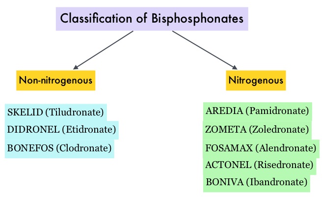
Bisphosphonates can also reduce bone pain and may improve Quality of life. Intravenous bisphosphonates, Pamidronate (AREDIA®) and Zoledronic acid (ZOMETA®) have been approved in the US for the treatment of bone metastases. Amino-bisphosphonate, ZOMETA® has however largely replaced AREDIA®, because of its superior efficacy. Both AREDIA® and ZOMETA® are administered IV every 3 to 4 weeks during the first year, following diagnoses of bone metastases. However, the optimal treatment schedule following this initial phase of treatment has remained unclear. Further, renal toxicity, long bone fractures and OsteoNecrosis of the Jaw (ONJ) have been identified as potential problems with bisphosphonate use.

CALGB 70604 (Alliance), is a randomized phase III study in which the efficacy of ZOMETA® administered every 4 weeks was compared with ZOMETA® administered every 12 weeks, in patients with breast cancer, prostate cancer or multiple myeloma, with bone metastases. In this non-inferiority trial, 1822 patients (Breast = 833, Prostate = 674, Myeloma= 270 and Other= 45) were randomly assigned 1:1, to receive ZOMETA® every 4 weeks or every 12 weeks for 2 years. 
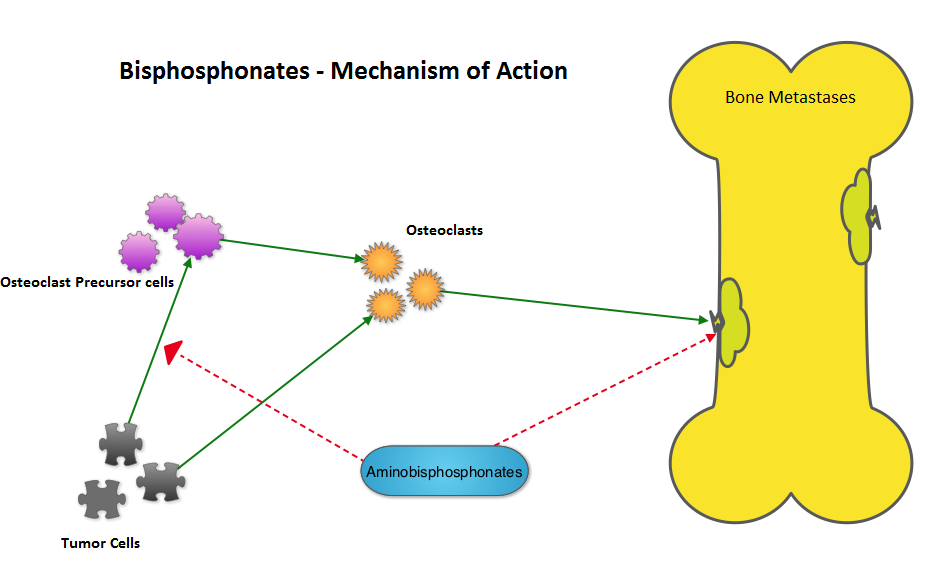
The primary endpoint was incidence of any Skeletal Related Event (SRE) and secondary endpoints included skeletal morbidity rates, performance status, pain using the Brief Pain Inventory and incidences of ONJ and renal dysfunction. Both treatment groups were well matched. Patients in this trial were stratified by disease and analyses by disease was pre-planned. It was noted that for the primary endpoint, there was no significant difference between the two treatment groups with 29% of patients in both treatment groups experiencing at least one SRE (P=0.79). With regards to secondary endpoints, there were still no significant differences between the two treatment groups, including renal dysfunction and ONJ. The authors pointed out that toxicities such as ONJ and renal dysfunction are more likely to occur after 2 years of treatment.

It was concluded that ZOMETA® administered every 3 months for 2 years is non-inferior to ZOMETA® administered every 4 weeks for 2 years, in patients with breast cancer, prostate cancer and multiple myeloma, with bone metastases. A less frequent dosing of ZOMETA® compared with the standard monthly dosing, may be more convenient for the patients and cost effective. CALGB 70604 (Alliance): A randomized phase III study of standard dosing vs. longer interval dosing of zoledronic acid in metastatic cancer. Himelstein AL, Qin R, Novotny PJ, et al. J Clin Oncol 33, 2015 (suppl; abstr 9501)