FDA Approves ARZERRA® Maintenance Therapy in Chronic Lymphocytic Leukemia

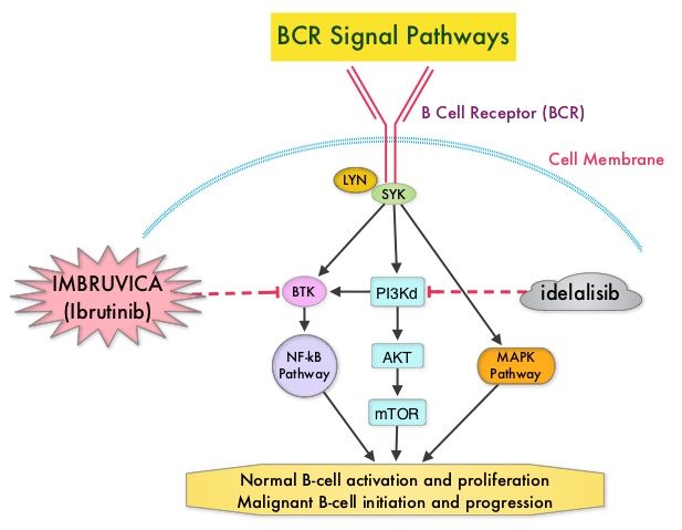
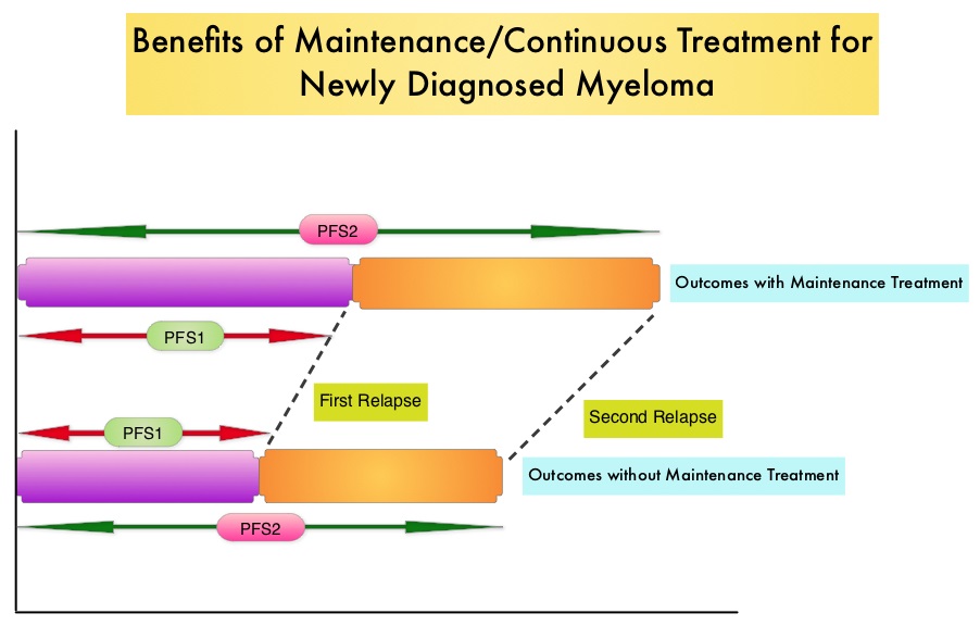
SUMMARY: The FDA on January 19, 2016 approved ARZERRA® (Ofatumumab) for extended treatment of patients who are in complete or partial response after at least two lines of therapy for recurrent or progressive Chronic Lymphocytic Leukemia (CLL). The American Cancer Society estimates that approximately 14,620 new cases of Chronic Lymphocytic Leukemia (CLL) were diagnosed in 2015 and approximately 4650 patients died from the disease. CLL is a disease of the elderly and the average age at the time of diagnosis is 72 years. ARZERRA® was previously approved for the treatment of treatment naive patients with CLL for whom FLUDARA® (Fludarabine) based therapy was considered inappropriate and also for patients with CLL refractory to FLUDARA® and CAMPATH® (Alemtuzumab). ARZERRA® is a second generation fully human IgG 1 monoclonal antibody. Unlike RITUXAN® (Rituximab), which is a chimeric monoclonal antibody, ARZERRA® targets a different region (different epitope) of the CD20 molecule. Monoclonal antibodies targeting CD20 destroy CD20 positive B cells by 3 different mechanisms. They include Antibody Dependent Cellular Cytotoxicity (ADCC), Complement Dependent Cytotoxicity (CDC) and programmed cell death (Apoptosis). Unlike RITUXAN®, ARZERRA® targets the small loop epitope of CD20 molecule which is proximal to the B cell membrane and this has been shown to be associated with highly efficient cell lysis through Complement Dependent Cytotoxicity. So, compared to RITUXAN®, ARZERRA® has stronger CDC, similar ADCC and does not appear to induce Apoptosis.

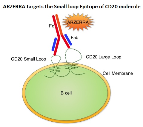
The PROLONG trial is an open-label, multicentre, randomised phase III study in which 474 patients with CLL whose disease had a complete or partial response after at least two lines of prior therapy, were randomly assigned in a 1:1 ratio to ARZERRA® (N=238) or observation (N=236). Patients in the ARZERRA® group received an initial dose of 300 mg given as an IV infusion followed by 1000 mg IV on Day 8. They subsequently received ARZERRA® 1000 mg IV every 8 weeks for up to 2 years. The median age was 65 years. The baseline characteristics in both treatment groups were well balanced. The primary endpoint was Progression Free Survival (PFS). Secondary endpoints included duration of response, Overall Survival, and safety. The median follow-up was 19•1 months. The median PFS with maintenance ARZERRA® was 29.4 months compared with 15.2 months in the observation group. This meant a 50% reduction in the risk of progression with maintenance ARZERRA® compared to observation (HR=0.50; P< 0.0001).

The most common adverse reactions in the ARZERRA® group were infusion reactions, neutropenia and upper respiratory tract infections. It was concluded that ARZERRA® is an important and new maintenance strategy in patients with relapsed CLL, to help delay disease progression. Ofatumumab maintenance versus observation in relapsed chronic lymphocytic leukaemia (PROLONG): an open-label, multicentre, randomised phase 3 study. van Oers MHJ, Kuliczkowski K, Smolej L, et al. The Lancet Oncology 2015: 16;1282-1284

XELOX Adjuvant Chemotherapy Regimen Improves Overall Survival in Stage III Colorectal Cancer

SUMMARY: The American Cancer Society estimates that approximately 133,000 new cases of ColoRectal Cancer (CRC) will be diagnosed in the United States in 2015 and close to 50,000 are expected to die of the disease. Adjuvant chemotherapy for patients with resected locally advanced, node-positive (stage III) colon cancer has been the standard of care since 1990s with improved Overall Survival noted after 6 months of bolus schedule of 5-Fluouracil (5-FU) and Leucovorin. Subsequently, the Multicenter International Study of Oxaliplatin/5-Fluorouracil/Leucovorin in the Adjuvant Treatment of Colon Cancer (MOSAIC) and National Surgical Adjuvant Breast and Bowel Project (NSABP) C-07 trials showed that the addition of ELOXATIN® (Oxaliplatin) to infusional 5- FU and Leucovorin (FOLFOX) or bolus 5-FU and Leucovorin (FLOX) significantly prolonged 3 year Disease Free Survival (DFS) when given in an adjuvant setting, in patients with stage II or III colon cancer. However, there was no significant Overall Survival (OS) benefit noted in the NSABP C-07 study.

XELOXA or NO16968 is a multinational, open-label, randomized phase III study, which only enrolled patients with stage III disease. Patients (N=1886) were randomly assigned to receive a more convenient XELOX regimen (N=944) or 5-FU and Leucovorin (N=942). The later regimen was considered the standard therapy when this trial was designed.The XELOX regimen consisted of a ELOXATIN® 130 mg/m2 given as a 2-hour IV infusion on day 1 and XELODA® (Capecitabine) 1,000 mg/m2 PO twice daily on days 1 to 14, of a 3 week cycle, for a total of eight cycles. The 5-FU/Leucovorin regimens could be either the Mayo Clinic or Roswell Park regimens. The Mayo Clinic regimen consisted of Leucovorin 20 mg/m2 and 5-FU 425 mg/m2 IV push on days 1-5, repeated every 4 weeks, for a total of six cycles. The Roswell Park regimen consisted of Leucovorin 500 mg/m2 given as a 2-hour infusion and 5-FU 500 mg/m2 IV push at 1 hour after the start of the Leucovorin infusion, repeated every week for six weeks, followed by a 2 week rest period, for a total of four 8-week cycles. The primary end point of this study was Disease Free Survival (DFS). Secondary end points were Overall Survival (OS), Relapse Free Survival (RFS) and safety.

The 3 year DFS data was published in 2011 and it was then noted that the addition of ELOXATIN® to oral Fluoropyrimidine, XELODA® improved DFS (HR=0.80; P=0.0045), similar to the previously published MOSAIC and NSABP C-07 trials. The authors in this publication reported the final efficacy data and biomarker analysis from the NO16968 trial comparing bolus 5-FU and Leucovorin with XELODA® plus ELOXATIN® (XELOX) in resected stage III colon cancer. The 7 year DFS rates were 63% and 56% in the XELOX and 5-FU/Leucovorin groups respectively (HR=0.80; P=0.004). The Overall Survival rates after a median follow up of 7 years were 73% and 67% in the XELOX and 5-FU/Leucovorin groups respectively (HR=0.83; P=0.04). It was noted that in the 498 patients who consented to the biomarker analysis, low tumor expression of DihydroPyrimidine Dehydrogenase was predictive for efficacy with XELOX regimen. There was however no statistically significant associations noted between any tumor biomarker and outcomes in the 5-FU/Leucovorin groups.

The authors concluded that in patients with resected stage III colon cancer, XELOX significantly improved Overall Survival compared to 5-FU/Leucovorin regimens and should be considered a standard adjuvant treatment option for patients with stage III disease. Tumor DihydroPyrimidine Dehydrogenase expression may be a clinically relevant biomarker for XELOX efficacy, but will require further evaluation. Capecitabine Plus Oxaliplatin Compared With Fluorouracil/Folinic Acid As Adjuvant Therapy for Stage III Colon Cancer: Final Results of the NO16968 Randomized Controlled Phase III Trial.Schmoll HJ, Tabernero J, Maroun J, et al. J Clin Oncol. 2015;33:3733-3740

Use of Biomarkers to Guide Decisions on Systemic Therapy for Women with Metastatic Breast Cancer American Society of Clinical Oncology Clinical Practice Guideline

SUMMARY: Breast cancer is the most common cancer among women in the US and about 1 in 8 women (12%) will develop invasive breast cancer during their lifetime. Approximately, 231,840 new cases of invasive breast cancer will be diagnosed in 2015 and over 40,000 women will die of the disease. The American Society of Clinical Oncology (ASCO) guidelines on the use of tumor markers in breast cancer are meant to provide evidence-based recommendations and guidance to practicing oncologists, on the appropriate use of breast tumor biomarker assays, for management of patients with metastatic breast cancer. These recommendations are for women with metastatic breast cancer being considered for systemic therapy or for changes in the drug or regimen they are receiving.

These guidelines were compiled after reviewing 17 clinical publications following an extensive literature search between 2006 and 2014. They included 11 studies that reported discordance in expression of hormone receptors or HER-2 between primary tumors and metastases, one randomized controlled study that addressed the use of a biomarker to decide whether to continue or change a treatment regimen and 5 prospective and retrospective studies that evaluated the clinical utility of biomarkers.

Should metastases be biopsied or otherwise sampled to test for changes from the primary tumor with respect to ER, PR, or HER2 status?

Patients with accessible, newly diagnosed metastases from primary breast cancer should be offered biopsy for confirmation of disease process and testing of ER, PR, and HER2 status. They should also be informed that if discordances are found, evidence is lacking to determine whether outcomes are better with treatment regimens based on receptor status in the metastases or the primary tumor. With discordance of results between primary and metastatic tissues, the Panel consensus is to preferentially use the ER, PR, and HER2 status from the metastasis to direct therapy, if supported by the clinical scenario and the patient's goals for care.

For women with metastatic breast cancer and with known ER, PR, and HER2 status, which additional tumor markers have demonstrated clinical utility to initiate systemic therapy or direct selection of a new systemic therapy regimen?

Decisions on initiating systemic therapy for metastatic breast cancer should be based on clinical evaluation, judgment, and patient preferences. There is no evidence at this time that initiating therapy solely on the basis of biomarker results beyond those of ER, PR, and HER2 improves health outcomes.

For women with metastatic breast cancer and with known ER, PR, and HER2 status, which additional tumor markers have demonstrated clinical utility to guide decisions on switching to a different drug or regimen or discontinuing treatment?

Recommendations for tissue biomarkers: In patients already receiving systemic therapy for metastatic breast cancer, decisions on changing to a new drug or regimen or discontinuing treatment should be based on clinical evaluation, judgment of disease progression or response, and the patient's goals for care. There is no evidence at this time that changing therapy based solely on biomarker results beyond ER, PR, and HER2 improves health outcomes, quality of life, or cost effectiveness.

Recommendations for circulating tumor markers: In patients already receiving systemic therapy for metastatic breast cancer, decisions on changing to a new drug or regimen or discontinuing treatment should be based on clinical evaluation, judgment of disease progression or response, and the patient's goals for care. There is no evidence at this time that changing therapy based solely on circulating biomarker results improves health outcomes, quality of life, or cost effectiveness. CEA, CA 15-3, and CA 27-29 may be used as adjunctive assessments to contribute to decisions regarding therapy for metastatic breast cancer. Data are insufficient to recommend use of CEA, CA 15-3, and CA 27-29 alone for monitoring response to treatment. As such, it is also reasonable for clinicians to not use these markers as adjunctive assessments.

For biomarkers shown to have clinical utility to guide decisions on systemic therapy for metastatic disease in questions 2 and 3, what are the appropriate assays, timing, and frequency of measurement?

Decisions for systemic therapy should be influenced by ER, PR, and HER2. ASCO recently updated the guideline addressing optimization of HER2 assays. To date, clinical utility has not been demonstrated for any additional biomarkers.

Poznak CV, Somerfield MR, Bast RC, et al. J Clin Oncol 2015;33:2695-2704

Direct Oral Anticoagulants Versus Vitamin K Antagonists in the Frail Elderly

SUMMARY: There are presently four Direct Oral AntiCoagulants (DOACs) approved in the United States for the treatment of Venous ThromboEmbolism. They include PRADAXA® (Dabigatran), which is a direct thrombin inhibitor and XARELTO® (Rivaroxaban), ELIQUIS® (Apixaban), SAVAYSA® (Endoxaban), which are Factor Xa inhibitors. Compared to Vitamin K Antagonist COUMADIN® (Warfarin), the Direct Oral AntiCoagulants have a rapid onset of action, wider therapeutic window, shorter half-lives (7-14 hours in healthy individuals), no laboratory monitoring and fixed dosing schedule. The half life of these agents can however be prolonged in those with renal insufficiency. The FDA in October, 2015, granted accelerated approval to PRAXBIND® (Idarucizumab), for those patients treated with PRADAXA® (Dabigatran), when reversal of the anticoagulant effects of PRADAXA® is needed for emergency surgery/urgent procedures, or in life-threatening or uncontrolled bleeding. Unlike bleeding caused by COUMADIN® which can be reversed using Vitamin K or Fresh Frozen Plasma, there are no specific agents presently available, for reversing bleeding caused by the other Direct Oral AntiCoagulants or for stopping the anticoagulant effects of these drugs, in patients who need urgent surgical intervention.

A recently published study has shown that the availability of Direct Oral AntiCoagulants has increased the number of doctor office visits, driven by new visits related to Direct Oral AntiCoagulant (DOAC) use in Atrial Fibrillation patients. It was noted that the utilization of DOAC’s was comparable to the use of COUMADIN® (Warfarin) for Atrial Fibrillation patients (Am J Med. 2015;128:1300-1305).

The discussion by the authors in this article, revolve around a 86-year old female weighing 55 kg, who presented to the ED with persistent Epistaxis. She had a history of Atrial Fibrillation and has been taking XARELTO® (Rivaroxaban) 20 mg, once daily, for stroke prevention for 1 year. She also had a history of peripheral arterial disease and was on Aspirin 80 mg, daily for primary prevention for 9 months. It was noted that the chromogenic anti–Factor Xa assay showed that her plasma concentration of XARELTO® was supratherapeutic. So, treatment with XARELTO® was discontinued. Nonetheless, two days later, the plasma XARELTO® concentration was still within the therapeutic range rather than subtherapeutic, suggesting slow drug elimination. The medical team discontinued her Aspirin and switched her from XARELTO® to COUMADIN® (Warfarin) and discharged her.

Several valuable recommendations made from this case are worth mentioning-

1) Older age and renal insufficiency are important factors contributing to bleeding while on DOACs. Because DOACs are partially excreted by the kidneys, dose reductions are recommended in the event of renal impairment. In a study evaluating the bleeding risk with PRADAXA® (Dabigatran) in the frail elderly, two thirds of patients were older than 80 years, and close to 60% of these patients had moderate or severe renal impairment.

2) Because patients with severe renal impairment have been excluded from phase III studies, DOACs should be avoided in this population along with those patients with extreme body weights.

3) Addition of Aspirin to oral anticoagulants is appropriate for up to 12 months after acute coronary syndromes, percutaneous coronary interventions, or stenting procedures. However, this combination therapy increases the risk of major bleeding by 50% compared with oral anticoagulant use alone.

4) For patients with mechanical heart valves, a combination of Aspirin plus oral anticoagulant may be appropriate. However, only a Vitamin K Antagonist (VKA) such as COUMADIN® should be used and DOACs are not recommended.

5) Although routine laboratory testing of patients taking a DOAC is not required, elderly patients should be closely monitored, with particular attention to renal function. A normal Prothrombin Time excludes supratherapeutic XARELTO® plasma levels, and XARELTO® levels can be measured using anti–Factor Xa chromogenic assays.

In conclusion, even though DOACs have several advantages compared with VKAs, DOACs should be avoided in frail elderly patients.

Optimizing the Safe Use of Direct Oral Anticoagulants in Older Patients – A Teachable Moment. Sennesael A, Dogné J and Spinewine A. JAMA Intern Med. 2015;175:1608-1609

IMBRUVICA® as Initial Therapy for Patients with Chronic Lymphocytic Leukemia

SUMMARY: The American Cancer Society estimates that approximately 14,620 new cases of Chronic Lymphocytic Leukemia (CLL) were diagnosed in 2015 and approximately 4650 patients died from the disease. CLL is a disease of the elderly and the average age at the time of diagnosis is 72 years. There are two main types of lymphocytes, B and T lymphocytes/cells. B-cell CLL is the most common type of leukemia in adults. Normal B-cell activation and proliferation is dependent on B-cell receptor (BCR) signaling. This signaling is also important for initiation and progression of B-cell lymphoproliferative disorders. Bruton's Tyrosine Kinase (BTK) is a member of the Tec family of kinases, downstream of the B-cell receptor and is predominantly expressed in B-cells. It is a mediator of B-cell receptor signaling in normal and transformed B-cells. Following binding of antigen to the B-Cell Receptor, kinases such as Syk (Spleen Tyrosine Kinase), Lyn (member of the Src family of protein tyrosine kinases) and BTK (Bruton's Tyrosine Kinase) are activated, with subsequent propagation through PI3K/Akt, MAPK, and NF-κB pathways. This results in B-cell activation and proliferation. IMBRUVICA® (Ibrutinib) is an oral, irreversible inhibitor of BTK and inhibits cell proliferation and promotes programmed cell death (Apoptosis) by blocking B-cell activation and signaling. The FDA initially granted accelerated approval to IMBRUVICA® in February 2014 for previously treated patients with CLL and this was followed by full FDA approval and a new treatment indication for high-risk CLL patients with 17p deletions, in July 2014.

In elderly CLL patients with comorbid conditions, Chlorambucil is often considered as a standard first-line therapy because of the higher rate of toxicities associated with FLUDARA® (Fludarabine) and TREANDA® (Bendamustine). RESONATE-2 is a international, open-label, randomized, phase III trial, in which the efficacy of two oral agents , IMBRUVICA® and Chlorambucil, were compared, in previously untreated elderly patients with CLL or Small Lymphocytic Lymphoma.

In this study, 269 treatment naïve patients with CLL or Small Lymphocytic Lymphoma, who were 65 years of age or older, were randomly assigned in a 1:1 ratio, to receive IMBRUVICA® 420 mg PO once daily (N=136) or Chlorambucil at a dose of 0.5 mg/kg on days 1 and 15 of each 28 day cycle, increased to a maximum of 0.8 mg/kg, if tolerated (N=133). Patients with chromosome del (17p) were excluded. The median age was 73 years and 70% of patients were over age 70. The primary end point was Progression Free Survival (PFS) and secondary end points included, Overall Response Rate (ORR), Overall Survival (OS), and sustained hematologic improvement.

With a median follow-up of 18.4 months, patients in the IMBRUVICA® group had a significantly longer Progression Free Survival (PFS) compared to the Chlorambucil group (median not reached versus 18.9 months), with a risk of progression or death 84% lower with IMBRUVICA®, compared to Chlorambucil (HR=0.16; P<0.001). IMBRUVICA® significantly prolonged Overall Survival, with an estimated Overall Survival rate of 98% at 24 months, compared to 85% with Chlorambucil, with a 84% reduction in the risk of death (HR=0.16; P=0.001). Further, IMBRUVICA® significantly improved Overall Response Rate, compared with Chlorambucil (86% vs 35%; P< 0.001) and also significantly improved hemoglobin and platelets levels from baseline values, compared with Chlorambucil. The most common adverse events associated with IMBRUVICA® were diarrhea, fatigue, cough and nausea and were mostly Grade 1 toxicities. The authors concluded that IMBRUVICA® significantly improves PFS, OS and ORR, compared to Chlorambucil, in previously untreated patients with CLL or Small Lymphocytic Lymphoma, and should be considered for all elderly patients who are not candidates for aggressive systemic therapy. Ibrutinib as initial therapy for patients with chronic lymphocytic leukemia. Burger JA, Tedeschi A, Barr PM, et al. N Engl J Med 2015; 373:2425-2437

Hepatitis B Antiviral Prophylaxis for Cancer Patients with Solid Tumors Receiving Chemotherapy

SUMMARY: The Centers for Disease Control and Prevention (CDC) estimates that there are 800,000 -1.4 million individuals with Chronic Hepatitis B (HBV) infection in the United States. Reactivation of HBV is a major concern in cancer patients who may be on chemotherapy or other immunosuppressive therapies, with the incidence of HBV reactivation ranging from 40%-60% in those who are positive for Hepatitis B surface antigen (HBsAg). HBV reactivation is preventable with prophylactic antiviral therapy, failing which it can result in delays in cancer treatment, as well as potentially fatal outcomes. Based on recently published data, showing the high risk for HBV reactivation among patients with hematological malignancies receiving B-cell-depleting agents such as RITUXAN® (Rituximab) or ARZERRA® (Ofatumumab), the FDA has urged health care providers to screen all patients for HBV infection, prior to starting therapy with these agents. HBV reactivation has been observed following chemotherapy for solid tumors, but the risk for reactivation in these settings has been unclear with insufficient evidence. The American Society of Clinical Oncology in 2010 rendered a Provisional Clinical Opinion (PCO), suggesting that there was insufficient evidence to determine the net benefits and harms of routine screening for HBV infection, in patients receiving chemotherapy and the recommendation was that screening be considered in those at increased risk for HBV infection or who receive highly immunosuppressive regimens.

This present study was conducted to determine the risk for HBV reactivation with and without antiviral prophylaxis and the benefit of prophylaxis in adults with solid tumors and chronic or resolved HBV infection. This meta-analysis included 26 original reports and the studies were independently reviewed by two investigators for study inclusion. HBV patients included in this study were receiving chemotherapy for any solid tumor with or without concomitant HBV prophylactic therapy. Study patients could receive long-term antiviral treatment or prophylaxis before chemotherapy initiation and the comparison was with those receiving chemotherapy without antiviral prophylaxis. The primary outcome was HBV reactivation as defined by a greater than 10-fold increase in HBV DNA levels from baseline or an absolute increase greater than 105 copies/mL in those with chronic HBV infection or the re-emergence of HBsAg when previously negative, in those with resolved HBV infection. Secondary outcomes included HBV-related hepatitis, interruption or delay in chemotherapy, acute liver failure with coagulopathy and hepatic encephalopathy and death.

It was noted that in patients with chronic HBV infection receiving chemotherapy, the risk for HBV reactivation without antiviral prophylaxis ranged from 4% to 68% (median, 25%). The risk for HBV reactivation, HBV-related hepatitis, and chemotherapy interruption was reduced by more than 80% with antiviral prophylaxis. Interestingly, in patients with resolved HBV infection receiving chemotherapy, there was still a risk of HBV reactivation, with this risk ranging from 0.3% to 9%. The authors in this meta-analysis addressed a very important question and concluded that the risk for HBV reactivation in patients with chronic HBV, on chemotherapy for solid tumors, is similar to the risk with other types of immunosuppressive therapy. Cancer patients should therefore be screened for HBV before chemotherapy is initiated for solid tumors and started on antiviral prophylaxis. Paul S, Saxena A, Terrin N, et al. Hepatitis B Virus Reactivation and Prophylaxis During Solid Tumor Chemotherapy: A Systematic Review and Meta-analysis. Ann Intern Med. 2016; 164:30-40.

Pacritinib for Myelofibrosis – A New JAK-2 Inhibitor with a Better Toxicity Profile

SUMMARY: Myelofibrosis is a MyeloProliferative Neoplasm (MPN) characterized by a ineffective hematopoiesis, progressive fibrosis of the bone marrow and potential for leukemic transformation. This stem cell disorder is Philadelphia Chromosome negative and manifestations include anemia, splenomegaly and related symptoms such as abdominal distension and discomfort with early satiety. Cytokine driven debilitating symptoms such as fatigue, fever, night sweats, weight loss, pruritus and bone or muscle pain can further impact an individual’s quality of life. Myelofibrosis can be primary (PMF) or secondary to Polycythemia Vera (PV) or Essential Thrombocythemia (ET). 
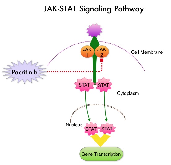
The JAK-STAT signaling pathway has been implicated in the pathogenesis of Myelofibrosis. This pathway normally is responsible for passing information from outside the cell through the cell membrane to the DNA in the nucleus, for gene transcription. Janus Kinase (JAK) family of tyrosine kinases are cytoplasmic proteins and include JAK1, JAK2, JAK3 and TYK2. JAK1 helps propagate the signaling of inflammatory cytokines whereas JAK2 is essential for growth and differentiation of hematopoietic stem cells. These tyrosine kinases mediate cell signaling by recruiting STAT’s (Signal Transducer and Activator of Transcription), with resulting modulation of gene expression. In patients with MPN, the aberrant myeloproliferation is the result of dysregulated JAK2-STAT signaling as well as excess production of inflammatory cytokines associated with this abnormal signaling. These cytokines contribute to the symptoms often reported by patients with MF. JAK2 mutations such as JAK2 V617F are seen in approximately 60% of the patients with PMF and ET and 95% of patients with PV. Unlike CML where the BCR-ABL fusion gene triggers the disease, JAK2 mutations are not initiators of the disease and are not specific for MPN. Further, several other genetic events may contribute to the abnormal JAK2-STAT signaling.

Pacritinib is a potent JAK2 inhibitor, without significant JAK1 inhibition. Preliminary studies have shown minimal myelosuppression with Pacritinib. JAKAFI® (Ruxolitinib) is a potent JAK1 and JAK2 inhibitor and is not safe for patients with low platelet counts. PERSIST-1 is a phase III study in which 327 patients with intermediate or high-risk Primary MyeloFibrosis (PMF), Post–Polycythemia Vera MF or Post–Essential Thrombocythemia MF were enrolled. Patients were randomized in a 2:1 ratio to receive Pacritinib 400 mg once daily (N=220) or Best Available Therapy (BAT) excluding JAKAFI® (N=107). Patients in the BAT group received Erythropoietin Stimulating Agents, Immunomodulatory drugs such as THALOMID® (Thalidomide), REVLIMID® (Lenalidomide) and Hydroxyurea. Because patients with very low platelet counts were enrolled in this study, JAKAFI® therapy was excluded, as JAKAFI® is not considered safe for patients with low platelet count. Approximately 32% of the patients had a platelet count of less than 100,000/µL and 15% had a platelet count of less than 50,000/ µL. About 75% of the patients were JAK2V617F positive. The median duration of treatment was 16.2 months in the Pacritinib group and 5.9 months in the BAT group. The primary endpoint was the proportion of patients achieving 35% or more reduction in the spleen volume at 24 weeks. Secondary endpoints included the proportion achieving 50% or more reduction in MyeloProliferative Neoplasm symptom score at 24 weeks.

At 24 weeks of treatment, 19.1% of patients in the Pacritinib group experienced 35% or more reduction in spleen volume compared to 4.7% in the Best Available Therapy (BAT) group (P=0.0003). This benefit was even more so in the subgroup of patients with the lowest platelet counts (less than 50,000/ µL), with 33.3% in the Pacritinib group demonstrating spleen volume reduction, compared to none in the BAT group. Patients in the Pacritinib arm were much more likely to experience more than a 50% reduction in symptoms, compared to BAT group at 24 weeks (24.5% vs 6.5%; P<0.0001), with significant improvements in fatigue, early satiety, abdominal discomfort, pruritus, night sweats and bone pain. This symptom improvement was noted by 4-8 weeks. Approximately 25% of the patients in the Pacritinib group achieved transfusion independence compared with none in the control group (P=0.043). The most common adverse events associated with Pacritinib were diarrhea, nausea and vomiting.

The authors concluded that Pacritinib significantly reduces spleen volume and Myelofibrosis associated symptoms, and fulfills an unmet need for Myelofibrosis patients with low platelet count, in addition to achieving RBC transfusion independence. Results of the PERSIST-1 phase III study of pacritinib (PAC) versus best available therapy (BAT) in primary myelofibrosis (PMF), post-polycythemia vera myelofibrosis (PPV-MF), or post-essential thrombocythemia-myelofibrosis (PET-MF). Mesa RA, Egyed M, Szoke A, et al. J Clin Oncol 33, 2015 (suppl; abstr LBA7006)

Continuous Therapy Significantly Improves Outcomes compared to Fixed Duration of Therapy in Patients with Newly Diagnosed Multiple Myeloma

SUMMARY: Multiple Myeloma is a clonal disorder of plasma cells in the bone marrow and the American Cancer Society estimates that in the United States, close to 27,000 new cases will be diagnosed in 2015 and 11,240 will die of the disease. Maintenance or Continuous Treatment in patients with newly diagnosed multiple myeloma following induction and consolidation, can result in significantly longer Progression Free Survival (PFS) and Overall Survival (OS), compared to those patients who receive therapy for a fixed duration of time. Not all studies however, have shown Overall Survival benefit. It has been hypothesized that Continuous Treatment could result in resistance to therapy which in turn could reduce the duration of subsequent remission after first relapse and negatively impact overall survival.To address this controversy, the authors conducted a pooled analysis of the outcomes of three randomized phase III trials, coordinated by the same principal investigator, designed to compare Continuous Treatment to Fixed Duration Therapy, in patients with newly diagnosed multiple myeloma.

In trial RV-MM-209, patients were randomized to either induction with Lenolidomide (REVLIMID®), followed by consolidation and subsequent maintenance with REVLIMID® (Continuous Treatment) or Fixed Duration Therapy which entailed REVLIMID® based induction followed by consolidation but no maintenance therapy. In the GIMEMA0305 trial, the randomization was between Bortezomib (VELCADE®) based induction followed by maintenance treatment (Continuous Treatment) and VELCADE® induction, with no maintenance treatment (Fixed Duration Therapy). In the CC-5013-MM-015 study, the comparison was between REVLIMID® in combination with Melphalan and Prednisone followed by REVLIMID® maintenance until disease progression and placebo given along with Melphalan and Prednisone.

The trial investigators assessed PFS1 as the time from diagnosis to the occurrence of 1st relapse, PFS2 as time from diagnosis to the occurrence of 2nd relapse and Overall Survival as time from diagnosis to death , incorporating the duration of both 1st and 2nd remission. They then evaluated PFS1, PFS2 and OS, in newly diagnosed multiple myeloma patients who received Continuous Therapy or Fixed Duration Therapy. In this pooled analysis of three trials, 604 patients were randomized to Continuous Treatment and 614 patients were randomized to Fixed Duration Therapy. Four hundred and seventeen (N=417) in the Continuous Therapy group and 410 patients in the Fixed Duration Therapy group were eligible for comparative analysis. The median follow up was 52 months.

Patients receiving Continuous Treatment had significantly prolonged PFS1 (median 32 months versus 16 months; HR=0.47; P<0.001), PFS2 (median 55 months versus 40 months; HR=0.61; P=0.001) and OS (4 year OS 69% versus 60%; HR=0.69; P=0.003), when compared with Fixed Dose Therapy. The authors evaluated the PFS and OS from first relapse to second relapse and from first relapse to death respectively, and they noted that the outcomes were similar among patients who received Continuous Treatment or Fixed Dose Therapy following initial diagnosis.

The authors concluded that Continuous Treatment significantly improved PFS1, PFS2, and OS and findings from this pooled analysis suggested that the clinical benefit observed during first remission was not negated by a shorter second remission and Continuous Treatment did not induce tumor resistance. Continuous Treatment may be essential, as patients with multiple myeloma will always have some residual disease. It should be noted that certain institutions including the Mayo Clinic cap Continuous/Maintenance treatment at approximately 2 years, due to the lack of randomized comparative data, on the value of prolonged maintenance beyond 2 years. Continuous Therapy Versus Fixed Duration of Therapy in Patients With Newly Diagnosed Multiple Myeloma. Palumbo A, Gay F, Cavallo F, et al. J Clin Oncol 2015;33:3459-3466

ACP Guidelines for Evaluation of Patients with Acute Pulmonary Embolism

SUMMARY: The Center for Disease Control and Prevention (CDC) estimates that approximately 1-2 per 1000 individuals develop Deep Vein Thrombosis/Pulmonary Embolism (PE) each year in the United States, resulting in 60,000 – 100,000 deaths. VTE is the third leading cause of cardiovascular mortality with a mortality rate of up to 25% in those with untreated acute Pulmonary Embolism. The American College of Physicians in 2015 released pulmonary embolism guidelines after noting that there has been a dramatic increase in the testing for suspected acute Pulmonary Embolism (PE). 
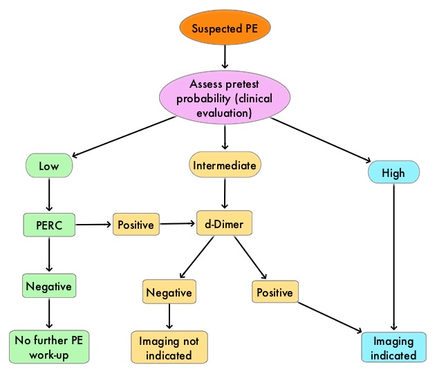
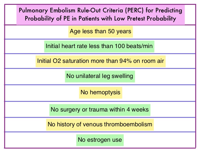
There has also been an overuse of CT imaging and plasma d-Dimer measurement, without improvement in care, but rather harming the patient and increasing expenditure. The validated clinical decision tools, in addition to physician’s clinical judgment include the Wells and Geneva Scoring System. The PERC (see table) criteria includes 8 elements, which if absent in low risk patients rules out PE. These practice guidelines were developed to provide practical advice, based on the best available evidence.

Best Practice Advice 1: Clinicians should use validated clinical prediction tools to estimate pretest probability, in patients in whom acute PE is being considered. These clinical decision tools include physician’s clinical judgment as well as Wells and Geneva Scoring System.

Best Practice Advice 2: The PERC (see table) criteria includes 8 elements. Clinicians should not obtain d-Dimer measurements or imaging studies in patients with a low pretest probability of PE and who meet all Pulmonary Embolism Rule-Out (PERC) Criteria.

Best Practice Advice 3: For patients who have an intermediate pretest probability of PE or in patients with low pretest probability of PE who do not meet all PERC Criteria, clinicians should obtain a high-sensitivity d-Dimer measurement as the initial diagnostic test in patients. Clinicians should not use imaging studies as the initial test in patients who have a low or intermediate pretest probability of PE.

Best Practice Advice 4: Clinicians should use age-adjusted d-Dimer thresholds (age × 10 ng/mL rather than a generic 500 ng/mL) in patients older than 50 years because, normal d-Dimer levels increase with age.

Best Practice Advice 5: Clinicians should not obtain any imaging studies in patients with a d-Dimer level below the age-adjusted cutoff.

Best Practice Advice 6: Clinicians should obtain imaging with CT pulmonary angiography (CTPA) in patients with high pretest probability of PE. Clinicians should reserve ventilation–perfusion scans for patients who have a contraindication to CTPA or if CTPA is not available. Clinicians should not obtain a d-Dimer measurement in patients with a high pretest probability of PE because, a negative d-Dimer test will not preclude the need for imaging.

Evaluation of Patients with Suspected Acute Pulmonary Embolism: Best Practice Advice From the Clinical Guidelines Committee of the American College of Physicians. Raja AS, Greenberg JO, Qaseem A, et al. Ann Intern Med. 2015;163:701-711

Adjuvant PROLIA® Significantly Reduces Fracture Risk for Breast Cancer Patients on Aromatase Inhibitors

SUMMARY: Breast cancer is the most common cancer among women in the US and about 1 in 8 women (12%) will develop invasive breast cancer during their lifetime. Approximately, 231,840 new cases of invasive breast cancer will be diagnosed in 2015 and over 40,000 women will die of the disease. Approximately 75% of patients with breast cancer are hormone receptor positive (Estrogen Receptor/Progesterone Receptor positive) and this is a predictor of response to endocrine therapy. These patients are often treated with anti-estrogen therapy as first line treatment. In premenopausal woman, the ovary is the main source of estrogen production, whereas in postmenopausal women, the primary source of estrogen is the Aromatase enzyme mediated conversion of androstenedione and testosterone to estrone and estradiol in extragonadal/peripheral tissues. Tamoxifen is a nonsteroidal Selective Estrogen Receptor Modulator (SERM) and works mainly by binding to the Estrogen Receptor and thus blocks the proliferative actions of estrogen on the mammary tissue. Anastrozole,Letrozole and Exemestane are Aromatase Inhibitors that bind reversibly to the aromatase enzyme and inhibit the conversion of androgens to estrogens in the extra-gonadal tissues. Aromatase inhibitors are associated with accelerated bone loss, leading to a decrease in Bone Mineral Density (BMD) and can thus cause osteopenia and osteoporosis, thereby increasing fracture risk. According to the WHO definitions, a healthy 30 year old adult (young adult) with the ideal Bone Mineral Density (BMD) is given a T-score of 0. A normal BMD is within 1 Standard Deviation-SD (+1 or −1) of the young adult mean. Osteopenia is between 1 and 2.5 SD below the young adult mean (−1 to −2.5 SD). Osteoporosis is 2.5 SD or more below the young adult mean (−2.5 SD or lower).

PROLIA® (Denosumab) is a monoclonal antibody that inhibits osteoclast formation, function and survival by selectively targeting the RANK ligand. In this randomized, double-blind, phase III trial, the authors evaluated the benefits of the anti-RANK ligand antibody PROLIA® (Denosumab) on bone health, in postmenopausal patients, with early stage hormone receptor-positive breast cancer, treated with Aromatase Inhibitors. Of the 3425 enrolled patients, 3420 patients were randomly assigned to receive PROLIA® 60 mg (N=1711) or placebo (N=1709) subcutaneously every 6 months. Majority of the patients participating in this study had breast cancer with good prognosis and only 25% of the patients required adjuvant chemotherapy. Patient received a median of 7 doses of PROLIA® . The primary endpoint was time from randomization to first clinical fracture.

Compared with placebo, PROLIA® significantly delayed time to first clinical fracture (HR=0.50; P<0•0001), with the PROLIA® group, half as likely to have a first clinical fracture as the placebo group. This benefit of lowering fracture risk was seen in subgroups of patients with BMD T-score of less than –1 as well as those with BMD T-score of -1 or greater. Further, patients in the PROLIA® group had improvements in BMD from baseline, of the lumbar spine, total hip, and femoral neck, compared to the placebo group, which showed worsening at all sites (P<0.0001). At 3 years, patients in the PROLIA® group had significantly lower risk of both new, or worsening vertebral fractures. No cases of osteonecrosis of the jaw bone were reported.

The authors concluded that PROLIA® administered in an adjuvant setting, significantly reduces the risk of fractures in postmenopausal women with breast cancer receiving Aromatase Inhibitors, without added toxicity. Adjuvant denosumab in breast cancer (ABCSG-18): a multicentre, randomised, double-blind, placebo-controlled trial. Gnant M, Pfeiler G, Dubsky PC, et al. The Lancet 2015;386:433-443