Management of Localized or Locally Advanced Oropharyngeal Squamous Cell Carcinoma – ASTRO Clinical Practice Guideline

SUMMARY: The American Cancer Society estimates that 49,670 people will be diagnosed with Oral cavity and Oropharyngeal cancer in 2017 and an estimated 9700 patients will die of the disease. Over 90% of these malignancies are Squamous Cell Carcinomas (SCCs). OroPharyngeal Squamous Cell Carcinoma (OPSCC) involves the tonsils and base of the tongue, and recent studies have shown that over 70% of these tumors are caused by Human Papilloma Virus (HPV) and HPV-16 is the predominant type present in the tumor cells. The CDC estimates that more than 2,370 new cases of Human Papilloma Virus associated OPSCC are diagnosed in women and nearly 9,356 are diagnosed in men, each year in the United States, and this incidence has been on the rise.

The American Society for Radiation Oncology (ASTRO) convened the OroPharyngeal Squamous Cell Carcinoma (OPSCC) Guideline Panel which consisted of a multidisciplinary team of radiation, medical, and surgical oncologists, to perform a systematic literature review of studies published from January 1990 through December 2014, in an attempt to present evidence-based guidelines, for the treatment of OPSCC, with definitive or adjuvant Radiation Therapy (RT).

The following are the key questions and recommendations of the Guideline panel.

(1) When is it appropriate to add systemic therapy to definitive RT in the treatment of OPSCC?

Stage IVA-B disease

A) Patients with stage IVA-B tumors receiving definitive RT should receive concurrent high-dose intermittent Cisplatin.

B) Patients who are medically unfit for high-dose Cisplatin should receive concurrent Cetuximab or Carboplatin-Fluorouracil.

C) Weekly Cisplatin may be considered for patients who are medically unfit for high-dose Cisplatin.

D) Concurrent Cetuximab should not be administered in combination with chemotherapy.

E) Intra-arterial chemotherapy should not be used in this patient group.

Stage III disease

A) Patients with T3 N0-1 tumors should receive concurrent systemic therapy.

B) Patients with T1-T2 N1 tumors who are at a significant risk for locoregional recurrence may be considered for concurrent systemic therapy.

Stage I to II disease

Concurrent systemic therapy is not recommended for this patient group as there is no evidence supporting the use of systemic therapy in this generally favorable population.

(2) When is it appropriate to deliver PostOperative RT (PORT) with and without systemic therapy following primary surgery for OPSCC?

A) Concurrent high-dose intermittent Cisplatin should be delivered with PORT to patients with positive surgical margins and/or extracapsular nodal extension, independent of HPV status or the extent of extranodal tumor.

B) Concurrent weekly Cisplatin may be administered with PORT to patients who are considered inappropriate for standard high-dose intermittent Cisplatin, after a careful discussion of patient preferences and the limited evidence supporting this treatment schedule.

C) For the high-risk postoperative patient unable to receive Cisplatin-based concurrent chemoradiation therapy, RT alone should be routinely delivered without concurrent systemic therapy. Given the limited evidence supporting alternative regimens, treatment with non-Cisplatin systemic therapy should be accompanied by a careful discussion of the risks and unknown benefits of the combination.

D) Patients treated with PORT should not receive concurrent weekly Carboplatin, weekly Docetaxel or Cetuximab, either alone or in combination with chemotherapy, although such regimens are currently under investigation.

E) Patients treated with PORT should not receive concurrent Mitomycin-C, alone or with Bleomycin, given the limited evidence and experience supporting its use.

F) Postoperative chemotherapy should not be delivered alone or sequentially with postoperative RT.

Intermediate-risk pathologic factors such as lymphovascular invasion (LVI), perineural invasion (PNI), T3-4 disease, or positive lymph nodes

A) These patients should not routinely receive concurrent systemic therapy with PORT.

B) Concurrent Cisplatin-based chemotherapy may be considered if the post operative pathologic findings suggest significant risk of locoregional recurrence.

C) PORT should be delivered to patients with pathologic T3 or T4 and pathologic N2 or N3 disease.

D) PORT may be delivered to patients with pathologic N1 disease after a careful discussion with the patient.

E) PORT may be delivered to patients with LVI and/or PNI as the only risk factor(s), after a careful discussion with the patient.

No pathologic risk factors

PORT may be delivered to patients without conventional adverse pathologic risk factors only if the clinical and surgical findings imply a particularly significant risk of locoregional recurrence and after a careful discussion with the patient.

(3) When is it appropriate to use induction chemotherapy in the treatment of OPSCC?

Induction Chemotherapy should not be routinely delivered to patients with OPSCC.

(4) What are the appropriate dose, fractionation, and volume regimens with and without systemic therapy in the treatment of OPSCC?

A) For patients with stage III-IV disease a dose of 70 Gy over 7 weeks should be delivered to gross primary and nodal disease.

B) The biologically equivalent dose of approximately 50 Gy in 2 Gy fractions or slightly higher should be delivered electively to clinically and radiographically negative regions at-risk for microscopic spread of tumor.

C) Altered fractionation should be used in patients with stage IVA-B disease treated with definitive RT, who are not receiving concurrent systemic therapy as well as patients with T3 N0–1 disease not receiving concurrent chemoradiation. Additionally, it may be considered for patients with T1–2 N1 or T2 N0 disease at high risk for recurrence.

D) When treating OPSCC with concurrent systemic therapy,, either standard or accelerated fractionation may be implemented.

Adjuvant PORT

Adjuvant PORT should be delivered to regions of microscopically positive primary site surgical margins and extracapsular nodal extension at 2 Gy/fraction once daily to a total dose between 60 and 66 Gy.

Early T-stage tonsillar carcinoma

A) Unilateral RT should be delivered to patients with well-lateralized (confined to tonsillar fossa) T1-T2 tonsillar cancer and N0-N1 nodal category.

B) Unilateral RT may be delivered to patients with lateralized (<1 cm of soft palate extension but without base of tongue involvement) T1-T2 N0-N2a tonsillar cancer, without clinical or radiographic evidence of extracapsular extension, after careful discussion of patient preferences and the relative benefits of unilateral treatment versus the potential for contralateral nodal recurrence and subsequent salvage treatment.

Radiation therapy for oropharyngeal squamous cell carcinoma: Executive summary of an ASTRO Evidence-Based Clinical Practice Guideline. Sher DJ, Adelstein DJ, Bajaj GK, et al. DOI: http://dx.doi.org/10.1016/j.prro.2017.02.002

PET-CT in Colorectal Cancer Patients with a Rising CEA Can Detect Occult Recurrent Disease Amenable to Curative Therapy

SUMMARY: ColoRectal Cancer (CRC) is the third most common cancer diagnosed in both men and women in the United States. The American Cancer Society estimates that approximately 135,430 new cases of ColoRectal Cancer will be diagnosed in the United States in 2017 and over 50,260 patients are expected to die of the disease. Patients with ColoRectal Carcinoma are often followed up with regular CEA measurements after curative surgical resection and a rising CEA may be the first sign of CRC recurrence and warrants further investigation. Approximately 50% of the patients with early-stage disease after surgical resection will relapse with metastatic disease, during the first 3 years of follow-up. They may present with synchronous disease defined as distant metastases occurring within 6 months, and metachronous disease defined as distant metastases occuring beyond 6 months, of the primary diagnosis of CRC. These patients with oligo-metastatic disease, when detected early, may be potentially curable.

The authors in this study sought to (a) evaluate the utility of PET-CT in detecting occult disease recurrence in patients with raised CEA and (b) establish the prognostic effects of early detection of disease recurrence in patients with CRC. This retrospective analysis screened1200 patients from 2004 to 2010, with a confirmed diagnosis of CRC, who on follow up after curative therapy underwent FDG PET-CT imaging, for an elevated CEA, after normal findings on conventional investigations. Patients who had already received treatment with curative intent for synchronous or metachronous oligo-metastatic disease, including surgical resection, radiofrequency ablation (RFA), and radical chemoradiation, were also included. An elevated CEA level was defined as more than 3 ng/mL in nonsmokers and more than 5 ng/mL in smokers. A minimum of clinical and radiological follow up for 12 months or histopathological confirmation, were required, to ascertain recurrent disease. Eighty eight (N=88) patients who underwent PET-CT imaging because of any clinical indication and met the eligibility criteria, were included in the study. The mean age of patients was 66 years and 59% were male.

Recurrent disease was confirmed in 64% of the patients within the 12 months after their FDG PET-CT scan and the PET scan was able to detect the site of subtle relapse. The sensitivity of PET-CT to detect recurrence was 88% and the specificity was 88% as well. Fifty five percent (55%) of the patients with PET-CT-detected relapsed disease were deemed eligible for further curative therapy of whom 70% went on to receive potentially curative therapy. The Positive Predictive Value and Negative Predictive Value for FDG PET-CT to predict recurrence were 93% and 80% respectively.

The median Time To Progression for patients who received potentially curative therapy for the PET-CT-detected recurrence was 8.8 months versus 2.2 months for the patients not treated with curative intent. The median Overall Survival was 39.9 months for those who received potentially curative treatment versus 15.6 months for those who did not receive curative therapy. The 5-year survival rate in the curative group was 36.8% versus 6.1% in the non-curative group (P <0.001).

The authors concluded that early use of FDG PET-CT in patients with rising CEA levels is a highly sensitive and specific tool for the detection of occult ColoRectal Cancer recurrence, and in more than 50% of these patients, recurrent disease may still be amenable to curative therapy, and long-term survival can be achieved in a subgroup of this patient population. Survival Outcomes in Asymptomatic Patients With Normal Conventional Imaging but Raised Carcinoembryonic Antigen Levels in Colorectal Cancer Following Positron Emission Tomography-Computed Tomography Imaging. Khana K, Athaudaa A, Aitken K, et al. The Oncologist 2016;21:1502-1508

Pegylated Form of Recombinant Hyaluronidase Significantly Improves Progression Free Survival in Metastatic Pancreatic Cancer

SUMMARY: The American Cancer Society estimates that in 2017, about 53,670 people will be diagnosed with pancreatic cancer in the United States and about 43,090 patients will die of the disease. Some important risk factors for pancreatic cancer include increasing age, obesity, smoking history, genetic predisposition, exposure to certain dyes and chemicals, heavy alcohol use and pancreatitis. The best chance for long term survival is complete surgical resection, although this may not be feasible in a majority of the patients, as they present with advanced disease at the time of diagnosis. Based on the National Cancer Data Base, the 5 year observed survival rate for patients diagnosed with exocrine cancer of the pancreas is 14% for those with Stage IA disease and 1% for those with Stage IV disease. The FDA approved ABRAXANE® ((Paclitaxel albumin-bound particles) for use in combination with GEMZAR® (Gemcitabine) for the first line treatment of patients with metastatic adenocarcinoma of the pancreas. This approval was based on the demonstration of improved Overall Survival (OS) and Progression Free Survival (PFS), in a multicenter, international, open-label, randomized trial (MPACT study), when compared to single agent GEMZAR®.

PEGPH20 is a PEGylated form of recombinant human Hyaluronidase, for the potential systemic treatment of tumors that accumulate Hyaluronan (HA). PEGPH20 is an enzyme that temporarily degrades Hyaluronan, a dense component of the tumor microenvironment that can accumulate in higher concentrations around certain cancer cells and potentially constrict blood vessels and there by impede treatment access to tumor tissue. It is estimated that 35% to 40% of patients with pancreatic cancer have high expression of Hyaluronan and this biomarker may predict response to PEGPH20.

HALO 202 (Halo 109-202) is a phase 2 multicenter, randomized clinical trial, in which PEGPH20 in combination with ABRAXANE® and GEMZAR® was compared with ABRAXANE® and GEMZAR® alone, in treatment naive patients with metastatic pancreatic carcinoma. In this study, following enrollment of 146 patients in the first stage of the trial, the study was placed on hold to address concerns regarding thromboembolic events, in the group receiving PEGPH20. The protocol was amended to exclude those at high risk for a thromboembolic event and prophylaxis with Low Molecular Weight Heparin was required. One hundred thirty-three patients (N=133) were enrolled into the second stage of the trial for a total of 279 patients. Patients enrolled in stage 2 received Low Molecular Weight Heparin at a starting dose of 40 mg/day or 1 mg/kg/day, to prevent thromboembolic events. PEGPH20 was administered at 3 µg/kg twice weekly for cycle 1 followed by weekly administration in subsequent cycles. ABRAXANE® and GEMZAR® were administered at their standard FDA-approved doses. Tumor biopsy samples for the Hyaluronan analysis were available for 138 patients treated with PEGPH20 and 79 patients treated in the control group across both stages of the study. Overall, 49 patients in the PEGPH20 arm and 35 in the control group had Hyaluronan expression of 50% or more. The primary endpoint of the study was Progression Free Survival (PFS) across the entire treatment group. Following change in the treatment protocol, a second primary endpoint was added to assess thromboembolic event rate. Secondary endpoints included Objective Response Rate, PFS by Hyaluronan level, and Overall Survival. The second stage of this study was also utilized to validate a companion diagnostic for Hyaluronan (HA) levels.

It was noted that across the overall study population, there was a statistically significant increase in Progression Free Survival (PFS) in patients with high levels of Hyaluronan (HA-High) treated with PEGPH20 plus ABRAXANE® and GEMZAR®, compared to HA-High patients receiving ABRAXANE® and GEMZAR® alone. Among the patients in the second stage of this study, there was a 91% improvement in median PFS for HA-High patients in the PEGPH20 group compared to the control group (8.6 months versus 4.5 months) and the additional primary endpoint of a reduction in the rate of thromboembolic events was achieved, in the PEGPH20 group. Across all patients, thromboembolic events were experienced by 14% of those in the PEGPH20 group versus 10% of those in the ABRAXANE® and GEMZAR® group. These events were lower in those receiving Low Molecular Weight Heparin at 1 mg/kg/day dose versus 40 mg/day (6% vs 10%, respectively). The most common adverse events were cytopenias.

The authors concluded that the addition of PEGPH20 to ABRAXANE® and GEMZAR® resulted in significant improvement in Progression Free Survival compared with ABRAXANE®/GEMZAR® alone, in treatment naïve patients with advanced pancreatic cancer. Patients with high levels of expression of the biomarker Hyaluronan, had the best outcomes suggesting that a biopsy-based biomarker for hyaluronan content can potentially identify patients who will have a meaningfully greater response when PEGPH20 is added to standard chemotherapy. A phase III study is underway, evaluating PEGPH20 in combination with ABRAXANE® and GEMZAR® in patients with metastatic pancreatic cancer, with high Hyaluronan levels. Countouriotis A. Study 202 Overall Results and Stage 2 Results [webcast]. Halozyme Investor Call; January 5, 2017. Final analysis of stage 1 data from a randomized phase II study of PEGPH20 plus nab-Paclitaxel/gemcitabine in stage IV previously untreated pancreatic cancer patients (pts), utilizing Ventana companion diagnostic assay. Bullock AJ, Hingorani SR, Wu XW, et al. J Clin Oncol 34, 2016 (suppl; abstr 4104)

Lower Dose of XARELTO® Adequate for Prevention of Recurrent Venous Thromboembolism

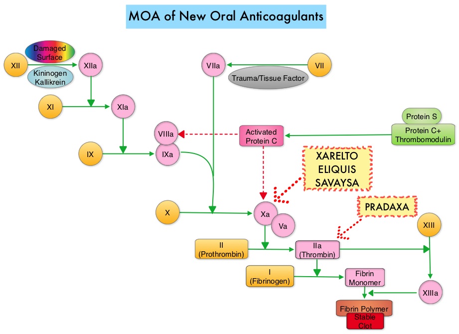
SUMMARY: The Center for Disease Control and Prevention (CDC) estimates that approximately 1-2 per 1000 individuals develop Deep Vein Thrombosis/Pulmonary Embolism (PE) each year in the United States, resulting in 60,000-100,000 deaths. Venous ThromboEmbolism (VTE) is the third leading cause of cardiovascular mortality, after myocardial infarction and stroke. With the exception of those with active malignancy, patients with VTE are often treated with Direct Oral Anticoagulant agents such as XARELTO® (Rivaroxaban) over vitamin K antagonists such as Warfarin because, Direct Oral Anticoagulant agents do not require routine laboratory monitoring or dose adjustment, have fewer interactions with food or other drugs and are associated with a lower risk of bleeding complications. Anticoagulation therapy is usually recommended for 3 months following an initial episode VTE in association with a transient risk factor such as surgery. In patients without reversible risk factors however, the risk of recurrent VTE is 10% or more during the first year, if anticoagulation therapy is discontinued. Extended anticoagulation therapy beyond 6 to 12 months even though effective is often not a common practice, for the fear of bleeding complications. Aspirin has been shown to reduce the risk of recurrent VTE when compared with placebo, but recurrence rate with this intervention is still significant at 5.1% per year.

The authors in this study compared the efficacy and safety of two doses of XARELTO® with Aspirin, in patients with Venous ThromboEmbolism, who had completed 6 to 12 months of anticoagulation therapy, and in whom there was uncertainty regarding the need for continued anticoagulation. In this randomized, double-blind, phase III trial, 3396 (N=3396) patients with Venous ThromboEmbolism were assigned, in a 1:1:1 ratio, to receive either XARELTO® 20 mg, XARELTO® 10 mg or Aspirin 100 mg given once daily. The primary efficacy outcome was symptomatic recurrent fatal or nonfatal Venous ThromboEmbolism, and the principal safety outcome was major bleeding. Secondary endpoints included whether the lower dose of XARELTO® was as effective as the higher dose and whether it was associated with less bleeding.

It was noted that after a median duration of treatment of 351 days, symptomatic recurrent fatal or nonfatal Venous ThromboEmbolism or unexplained death occurred in 1.5% of the patients assigned to XARELTO® 20 mg and 1.2% of the patients assigned to XARELTO® 10 mg compared with 4.4% among the patients assigned to the Aspirin group (P<0.001). Major or clinically relevant non-major bleeding occurred in 3.3%, 2.4%, and 2.0% of the patients, respectively.

It was concluded that among patients with Venous ThromboEmbolism for whom continued anticoagulation is a consideration, after an initial treatment course at usual therapeutic doses, the risk of a recurrent thromboembolic events were significantly lower with XARELTO® given at either a therapeutic dose (20 mg) or a prophylactic dose (10 mg), compared with Aspirin. There was no significant increase in bleeding rates. Rivaroxaban or Aspirin for Extended Treatment of Venous Thromboembolism. Weitz JI, Lensing AW, Prins MH, et al. for the EINSTEIN CHOICE Investigators. N Engl J Med 2017; 376:1211-1222

Sequencing Therapies in Metastatic Castrate Resistant Prostate Cancer

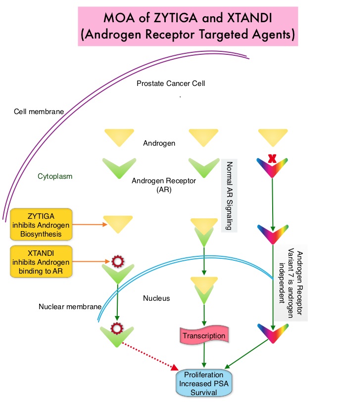
SUMMARY: Prostate cancer is the most common cancer in American men with the exclusion of skin cancer and 1 in 7 men will be diagnosed with prostate cancer during their lifetime. It is estimated that in the United States, about 161,360 new cases of prostate cancer will be diagnosed in 2017 and 26,730 men will die of the disease. The development and progression of prostate cancer is driven by androgens. Androgen Deprivation Therapy (ADT) has therefore been the cornerstone of treatment of advanced prostate cancer and is the first treatment intervention for hormone sensitive prostate cancer. Chemotherapy is usually considered for patients who progress on hormone therapy (Castrate Resistant Prostate Cancer-CRPC) and TAXOTERE® (Docetaxel) has been shown to improve Overall Survival (OS) of metastatic prostate cancer patients, who had progressed on Androgen Deprivation Therapy. Tumors in patients with CRPC are not androgen independent and continue to rely on Androgen Receptor signaling. Two oral Androgen Receptor Targeted Agents (ARTA) are presently available for metastatic CRPC. They include ZYTIGA® (Abiraterone) and XTANDI® (Enzalutamide). ZYTIGA® inhibits CYP 17A1 enzyme thus decreasing androgen biosynthesis and depletes adrenal and intratumoral androgens. XTANDI® competes with Testosterone and Dihydrotestosterone and avidly binds to the Androgen Receptor (AR), thereby inhibiting AR signaling and in addition inhibits translocation of the AR into the nucleus and thus inhibits the transcriptional activities of the AR. There is presently very little guidance with regards to the sequencing of these two oral agents.

It has remained unclear when a patient should receive chemotherapy following progression on AR-targeted therapies. To determine the ideal second line therapy in this patient population, the authors conducted a retrospective study, to assess if second line chemotherapy was associated with improved outcomes, compared with second line alternative AR Targeted Agents, in patients with a very short duration of response to first line Androgen Receptor Targeted Agents, in the US community oncology setting.

Using Altos electronic medical records, the authors identified 345 patients with metastatic CRPC who did not respond to first-line AR Targeted Agents (ZYTIGA® or XTANDI®) who then went on to receive second-line TAXOTERE® – Docetaxel or JEVTANA® – Cabazitaxel (chemotherapy cohort, N=147), or an alternative AR Targeted Agent (ARTA cohort, N=198), from May 2011 to Oct 2014. Patients receiving chemotherapy as second-line treatment, compared to those receiving second-line ARTA, were younger (median age, 74 vs 79 years) and had several poor prognostic factors including a higher mean PSA, LDH and Alkaline Phosphatase, as well as lower mean hemoglobin and increased opioid use. Treatment outcomes were evaluated from start of second-line treatment and second-line chemotherapy was compared to second-line ARTA. The primary endpoints included Prostate Specific Antigen (PSA) response (50% or more reduction) and Overall Survival (OS).

It was noted that more patients in the chemotherapy group had a PSA response compared to the AR Targeted Agent group (P=0.005), and there was a non-statistically significant trend toward improved Overall Survival for second-line chemotherapy versus AR Targeted Agent (adjusted HR=0.81; P=0.15). Among patients with poor prognostic features, those in the chemotherapy cohort had significantly improved Overall Survival (adjusted HR=0.55; P=0.009) compared with those in the AR Targeted Agent cohort.

The authors concluded that patients who do not respond well to first-line Androgen Receptor Targeted Agent and have poor prognostic features, should not receive a second AR-targeted agent but instead receive second-line chemotherapy, as this may confer a survival benefit. Real-world outcomes in patients with metastatic castration-resistant prostate cancer receiving second-line chemotherapy vs alternative androgen receptor-target agents (ARTA) after lack of response to first-line ARTA in US community oncology practices. Oh WK, Cheng WY, Miao R, et al. J Clin Oncol. 2017;35 (suppl 6S; abstr 214).

Restricting Indoor Tanning Can Impact Melanoma Incidence and Treatment Costs

SUMMARY: It is estimated that in the US, about 87,110 new cases of melanoma will be diagnosed in 2017 and about 9,730 patients will die of the disease. The incidence of melanoma has been on the rise for the past three decades. A major risk factor for most skin cancers is exposure to UltraViolet (UV) radiation, which damages the DNA of skin cells. The main source of UV rays is sunlight, tanning lamps and tanning beds. UV exposure also can cause cataracts and ocular melanoma. Indoor tanning, exposes individuals to both UVA and UVB rays and is particularly dangerous for those who begin indoor tanning during adolescence or early adulthood, putting them at a higher risk of developing melanoma. The association between indoor tanning beds, including those with the newer tanning technology, and melanoma, has been well established with a 22-34% increase in the risk of developing melanoma with indoor tanning. The International Agency for Research on Cancer has classified indoor tanning as a Class I carcinogen, based on its significant association with malignant melanoma. Indoor tanning with resulting exposure to ultraviolet radiation is a potentially modifiable behavior. According to the 2015 Youth Risk Behavior Surveillance System, approximately 15% of the high school teens are indoor tanning and according to the 2010 National Health Interview Survey, indoor tanners tended to be young, non-Hispanic white women of whom 32% are aged 18 to 21 years.

The authors in this study estimated the health and economic benefits of reducing indoor tanning in the United States. Using a statistical model that takes into consideration risk, that continues over time (Markov model), the authors estimated the number of melanoma cases and deaths that could be prevented and melanoma treatment costs saved, by reducing indoor tanning among minors younger than 18 years. These estimations were based on a cohort of 61.2 million individuals aged 14 years or younger in the United States and the probability of these individuals using indoor tanning, based on data from the 2013 Youth Risk Behavior Survey. The prevalence of indoor tanning was based on a recent meta-analysis.

The researchers noted that restricting the use of indoor tanning among minors younger than 18 years was estimated to prevent 61,839 cases of melanoma (4.9% reduction) and 6,735 melanoma deaths (4.7% reduction), over the lifetime of this group of individuals. The treatment cost-savings from these reductions was estimated to be $342.9 million over the lifetime of these individuals. These health and economic benefits increased as indoor tanning was further reduced.

The authors concluded that reducing indoor tanning has the potential to reduce melanoma incidence, mortality, and treatment costs, emphasizing the importance of continued public health efforts to identify and implement effective strategies to prevent melanoma. The potential impact of reducing indoor tanning on melanoma prevention and treatment costs in the United States: An economic analysis. Guy GP, Zhang Y, Ekwueme DU, et al. Journal of the American Academy of Dermatology 2017;76:226-233

Reduced Lung-Cancer Mortality with Low-Dose Computed Tomographic Screening

SUMMARY: The rationale for Lung Cancer screening is based on the National Lung Cancer Screening Trial (NLST) in which the use of low dose CT scan in high risk, healthy patients, resulted in a 20% reduction in lung cancer mortality, compared to screening with a chest x-ray. It is important that eligible people who are smokers participate in a smoking cessation program and quit smoking. Further, those eligible for screening should understand the limitations associated with any screening methodology and potential risks associated with procedures that may follow a false positive result.

Lung cancer screening is performed using a non-contrast low dose CT scan. Criteria for lung cancer screening include-

1) People 55-74 years of age with no signs or symptoms of lung cancer

2) Current or former smoker with a 30 pack year smoking history (Number of years smoked multiplied by the number of packs of cigarettes per day)

3) Current smokers are strongly urged to enter a smoking cessation program

4) Former smokers must have quit smoking within the past 15 years

People with serious comorbid conditions, those on home oxygen and individuals with metallic devices or implants in the chest or back (which can interfere with the scan) should be excluded from lung cancer screening. Lung cancer screening with low dose CT scan is presently not covered by most insurance plans. The National Lung Screening Trial Research Team. N Engl J Med 2011; 365:395-409

CABOMETYX® Superior to SUTENT® in Metastatic Renal Cell Carcinoma

SUMMARY: The American Cancer Society estimates that about 63,990 new cases of kidney cancer will be diagnosed in the United States in 2017 and about 14,400 patients will die from this disease. The VHL (Von Hippel-Lindau) protein is a tumor suppressor gene which is frequently mutated and inactivated in approximately 90% of clear cell Renal Cell Carcinomas (RCC). The VHL gene under normal conditions binds to Hypoxia-Inducible Factor (HIF-1 alpha) and facilitates degradation of this factor. Under hypoxic conditions and in patients having biallelic loss of function and mutation of VHL genes, HIF-1alpha is not degraded. Build up of HIF-1 alpha results in increased angiogenesis, increased tumor cell proliferation and survival, as well as metastasis.

SUTENT® (Sunitinib) is the standard first-line intervention for treatment naïve patients with advanced Renal Cell Carcinoma. VOTRIENT® (Pazopanib) another VEGFR-targeted therapy, is an alternative choice, as it was found to be non-inferior to SUTENT® in the COMPARZ trial. CABOMETYX® (Cabozantinib) is an oral, small-molecule Tyrosine Kinase Inhibitor (TKI) but, unlike SUTENT® which targets the Vascular Endothelial Growth Factor Receptors (VEGFR), CABOMETYX® additionally inhibits the action of tyrosine kinases MET and AXL. Both MET and AXL are up-regulated in Renal Cell Carcinoma as a consequence of VHL inactivation and increased expression of MET and AXL is associated with tumor progression and development of resistance to VEGFR inhibitors. CABOMETYX® was approved by the FDA in 2016 for the treatment of advanced Renal Cell Carcinoma (RCC), in patients who have received prior anti-angiogenic therapy.

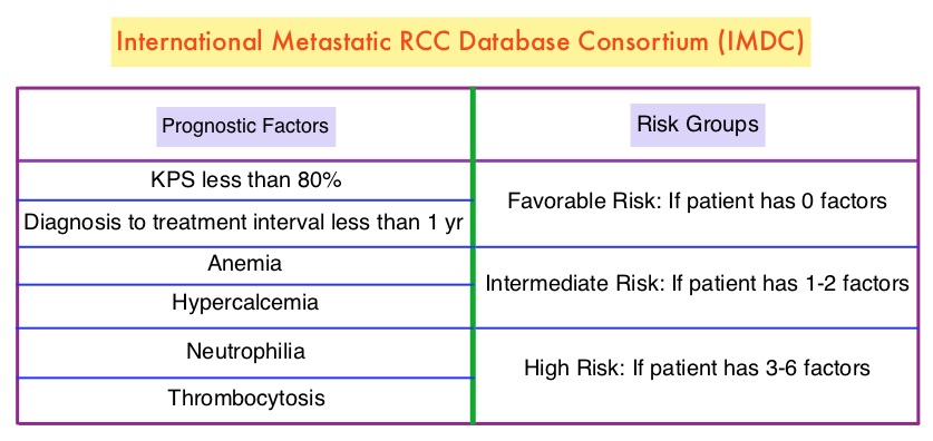
The Alliance for Clinical Trials in Oncology reported the results of a randomized, multicenter, open-label phase II CABOSUN trial, which compared CABOMETYX® with standard-of-care SUTENT®, in IMDC intermediate and poor-risk untreated patients with locally advanced or metastatic clear-cell RCC. The study population had a high rate of bone metastases, a known negative prognostic factor in RCC. This study enrolled 157 patients who were randomized in a 1:1 ratio to receive CABOMETYX® 60 mg orally daily (N=79) or SUTENT® 50 mg orally 4 weeks on, 2 weeks off (N=78). A treatment cycle was defined as 6 weeks in both study groups and treatment was continued until disease progression or intolerance to therapy. Patients were stratified by IMDC risk category (intermediate or poor) and presence of bone metastases. Crossover between treatment groups was not allowed. The median age was 63 years and 81% of the enrolled patients were classified as IMDC intermediate risk and 19% as poor risk, 36% of patients had bone metastases and 75% of the patients had prior nephrectomy. The Primary end point was Progression Free Survival (PFS) and Secondary end points included Objective Response Rate (ORR), Overall Survival (OS) and safety.

It was noted that the treatment with CABOMETYX® significantly increased median PFS compared with SUTENT® (8.2 versus 5.6 months) and was associated with a 34% reduction in rate of disease progression or death (HR=0.66; P=0.012). The ORR was 46% in the CABOMETYXreg; group compared with 18% in the SUTENT® group. The median OS with CABOMETYX® was 30.3 months versus 21.8 months with SUTENT® (HR=0.80), with this preliminary data showing a 20% decrease in the risk of death with CABOMETYX®. Grade 3 or 4 adverse events were similar in both treatment groups with more hypertension and Palmar-Plantar Erythrodysesthesia in the CABOMETYX® group whereas the SUTENT® group experienced more fatigue and hematologic toxicities. Discontinuation rates related to adverse events were similar in both treatment groups.

The authors concluded that CABOMETYX® significantly improved PFS and ORR compared to SUTENT®, in the initial treatment of patients with intermediate or poor-risk clear cell metastatic Renal Cell Carcinoma. CABOMETYX® is the first agent to demonstrate clinical superiority over SUTENT®, which has been the established standard of care for more than 10 years. The authors attributed the superiority of CABOMETYX® over SUTENT® due to its mechanism of action, which targets MET and AXL, in addition to VEGFR. Cabozantinib Versus Sunitinib As Initial Targeted Therapy for Patients With Metastatic Renal Cell Carcinoma of Poor or Intermediate Risk: The Alliance A031203 CABOSUN Trial. Choueiri TK, Halabi S, Sanford BL, et al. DOI: 10.1200/JCO.2016.70.7398 Journal of Clinical Oncology 35, no. 6 (February 2017) 591-597.

Location of Primary Tumor in the Colon Predicts Survival and Choice of Treatment in Metastatic Colorectal Cancer

SUMMARY: ColoRectal Cancer (CRC) is the third most common cancer diagnosed in both men and women in the United States. The American Cancer Society estimates that approximately 135,430 new cases of ColoRectal Cancer will be diagnosed in the United States in 2017 and over 50,260 patients are expected to die of the disease. Several published studies have demonstrated a nearly 20% reduced risk of death for patients diagnosed with left sided colon cancer compared with those who had right sided tumors. Venook and colleagues had previously presented their findings from a retrospective evaluation of the phase III 80405 clinical trial which included data from 1,025 metastatic ColoRectal Cancer patients with KRAS wild-type disease. The researchers assessed the impact of tumor location on Overall and Progression Free Survival in this group of patients. The median age was 59 yrs and 293 patients had a right-sided primary tumor (location in the cecum to hepatic flexure) and 732 patients had a left-sided primary tumor (location in the splenic flexure to rectum). It was noted that patients with tumors originating in the right side of the colon had much shorter median Overall Survival (19.4 months) compared to patients with left-sided tumors (33.3 months), (HR=1.60; P<0.001), with a 14 month survival improvement in the left versus right-sided primary tumors, for patients with metastatic disease. Tumor location in the colon also had a bearing on response to ERBITUX® (Cetuximab) and AVASTIN® (Bevacizumab). For the patient group who received ERBITUX®, the median Overall Survival (OS) was 36 months for patients with left-sided tumors but only 16.7 months for patients with right-sided tumors. For those who received AVASTIN®, the median OS was 31.4 months for patients with left-sided tumors and 24.2 months for those with right-sided tumors. The median Progression Free Survival was also influenced by the site of the primary tumor and was 11.5 months for left-sided tumors versus 8.9 months for right-sided tumors in the overall cohort of patients (HR = 1.26; P = 0.002). It was concluded that the biology of tumors originating in the right colon may be different from those originating in the left colon and regardless of KRAS mutational status, AVASTIN® based, first line chemotherapy regimen should be considered, for all patients with metastatic colorectal cancer, with a right-sided colon primary tumor and ERBITUX® should be avoided in this patient group.

Understanding that there is a difference in outcomes with biologic therapy, based on tumor location in the colon, the authors in this study evaluated the molecular variations of tumors arising in different parts of the left colon. Using protein expression, gene amplification and NextGen sequencing, 1,457 primary tumors (125 from splenic flexure to descending colon, 460 in the sigmoid colon and 872 in the rectum) were examined. MicroSatellite Instability (MSI) was measured by PCR and tumor mutational load was calculated using somatic nonsynonymous missense mutations.

They noted that the incidence of  to MSI significantly decreased from right colon (22%), to descending colon (7%),sigmoid colon (4%) to rectum (1%) and this was statistically significant (P=0.01). Rectal tumors had a higher frequency of TP53 and APC gene mutations and higher expression of TOPO1, ERCC1 and MGMT, compared to tumors in the descending colon. Compared to sigmoid colon tumors, rectal tumors had a higher expression of TOPO1, MGMT, TLE3 and TUBB3.

The authors concluded that tumors arising in the rectum have a distinct set of genetic alterations compared with other left colon tumors, and these distinct biologic entities may need to be addressed with individually tailored therapy. Molecular variances between rectal and left-sided colon cancers. Marshall J, Lenz H, Xiu J, et al. J Clin Oncol 35, 2017 (suppl 4S; abstract 522)