Gut Bacteria May Promote Pancreatic Cancer by Inducing Immune Suppression

SUMMARY: The American Cancer Society estimates that in 2018, about 55,440 people will be diagnosed with pancreatic cancer and about 44,330 people will die of the disease. Pancreatic cancer is the fourth most common cause of cancer-related deaths in the United States and Western Europe. Unfortunately, unlike other malignancies, very little progress has been made and outcomes for patients with advanced pancreatic cancer, has been dismal. Diagnosis is often made late in the course of the disease, as patients are often asymptomatic and early tumors cannot be detected during routine physical examination. Research has been underway evaluating the role of modifiable risk factors, early screening biomarkers, and tumor microenvironment and their influence on outcomes.

Recent published studies have shown the influence of gut microbiome alterations, in the carcinogenesis of pancreatic cancer. A commensal microbiome in a healthy individual maintains a symbiotic relationship conferring protection by its inflammatory-modulating activity, detoxification, hormonal homeostatic and metabolic effects of bacterial metabolites. An imbalanced microbiome can result in dysbiosis, and microbiome alteration has been reported to contribute to carcinogenesis of multiple malignancies. One classic example of influence of microbiome alteration contributing to carcinogenesis is Helicobacter pylori (H. pylori). Eradication of H. pylori causes regression of MALT lymphoma and decreases risk of metachronous gastric carcinoma after endoscopic resection for early stage gastric cancer.

An abundance and alteration of certain microbiomes has been shown to suppress monocytic cellular differentiation in pancreatic cancer leading to T-cell anergy. Targeting the microbiome and bacterial ablation has been shown to be associated with immunogenic reprogramming of the pancreatic tumor microenvironment, by reducing the myeloid-derived suppressor cells and increasing macrophage differentiation. Additionally, bacterial ablation also upregulates PD-1 expression, and enables the efficacy for checkpoint-based immunotherapy and reverses intratumoral immune tolerance. These findings have led to the conclusion that, endogenous microbiota promote immune suppression, characteristic of pancreatic ductal adenocarcinoma, and targeting microbiome potentially can modulate disease progression.

The present study was based on preclinical findings that cancerous pancreas harbors a population of bacteria that is a 1000 fold more, compared with normal pancreas in both mice and humans, and these select bacteria are differentially increased in the tumorous pancreas compared with the gut. The authors in this study noted that bacteria that are more abundant in pancreatic cancers include proteobacteria, actinobacteria, and fusobacteria species. These bacteria release cell membrane components such as lipopolysaccharides and proteins such as flagellins that shift macrophages into immune suppression and prevent the immune system from attacking tumor cells. Their study showed that eliminating these bacteria using antibiotics restored the ability of immune cells to recognize cancer cells, slowed pancreatic tumor growth, and reduced the cancer cell tumor burden by 50% in study animals. Specifically, it was noted that eradicating these bacteria from the gut and pancreas, by treating mice with antibiotics, slowed cancer growth and allowed the recognition of tumor cells by the immune system. Oral antibiotics also increased the efficacy of checkpoint inhibitors roughly 3 fold, thereby strongly improving antitumor immunity.

The researchers further pointed out that even though alterations in genes such as KRAS can result in abnormal cell growth and development of pancreatic cancer, the present study showed that bacteria can change the immune environment around cancer cells and facilitate rapid tumor growth in some patients more so than others, despite their similar genetic alterations. The authors hypothesized that changes in the genes that cause abnormal cell growth in the pancreas might also change the immune response, favoring the growth of different bacterial species, other than those found in healthy individuals. Environmental factors like diet, other medical conditions, or common medications, might also cause bacterial changes in the gut, that influence the pancreatic microbiome.

It was concluded that in pancreatic cancer, a distinct and abundant group of bacteria provide an immune suppressed environment, and addition of antibiotics improved the efficacy of a checkpoint inhibitor, in a mouse model of pancreatic ductal adenocarcinoma, as demonstrated by an increase in T cells that could attack the tumor. Studies are planned to evaluate the role of antibiotic combinations such as Ciprofloxacin and Metronidazole and their benefit in improving the efficacy of PD-1 inhibitors, in patients with pancreatic ductal adenocarcinoma. The Pancreatic Cancer Microbiome Promotes Oncogenesis by Induction of Innate and Adaptive Immune Suppression. Pushalkar S, Hundeyin M, Daley D, et al. DOI: 10.1158/2159-8290.CD-17-1134

ASTRO Issues New Updated Clinical Guideline for Whole Breast Radiation Therapy

SUMMARY: Breast cancer is the most common cancer among women in the US and about 1 in 8 women (12%) will develop invasive breast cancer during their lifetime. Approximately 266,120 new cases of invasive breast cancer will be diagnosed in 2018 and about 40,920 women will die of the disease. When it comes to adjuvant radiation therapy in breast cancer, the standard of care has been Whole Breast Irradiation (WBI), administered in Conventional Fractions (CF-WBI) of 180 to 200 cGy daily, to approximately 4500 to 5000 cGy, with or without a tumor bed boost. More recently, with clinical trials supporting the safety and effectiveness of HypoFractionated Whole Breast Irradiation (HF-WBI), the American Society for Radiation Oncology (ASTRO) task force, has issued a new clinical guideline for the use of whole-breast radiation therapy for breast cancer, which replaces the existing ASTRO Whole Breast Irradiation guideline published in 2011. With HypoFractionated Whole Breast Irradiation, patients receive larger doses of radiation over a shorter period of time, typically completing treatment in 3-4 weeks, compared with 5-7 weeks for CF-WBI.

The guideline recommendations were based on a systematic literature review, between January 2009 and May 2016, and created using ASTRO-approved tools, for grading evidence quality and recommendation strength. The ASTRO convened a task force to address 5 key questions focused on dose-fractionation for WBI, indications and dose fractionation for tumor bed boost, and treatment planning techniques for WBI and tumor bed boost. The recommendations are summarized below.

Whole-Breast Irradiation (without irradiation of regional lymph nodes) – Delivery and Dosing

1) Treatment decisions, including radiation techniques (Hypofractionated versus Conventional Fractions) should be a shared decision between patient and physician and should be individualized to each patient.

2) For women with invasive breast cancer receiving WBI with or without inclusion of the low axilla, the preferred dose-fractionation scheme is HF-WBI to a dose of 4000 cGy in 15 fractions or 4250 cGy in 16 fractions.

3) The decision to offer HF-WBI should be independent of age, tumor grade, hormone receptor status, HER2 receptor status, surgical margin status, breast size, breast cancer laterality, chemotherapy received prior to radiation and trastuzumab or endocrine therapy received prior to or during radiation.

4) HF-WBI may be used as an alternative to CF-WBI in patients with DCIS.

5) CF-WBI may be preferred over HFWBI when treating primary breast cancers with rare histologies that are most commonly treated with CF when arising in other parts of the body.

6) In patients with breast augmentation, either HF-WBI or CF-WBI may be used.

Tumor Bed Boost

1) A tumor bed boost is recommended for patients with invasive breast cancer 50 years or younger with any grade tumor, age 51-70 years with high grade tumor, or a positive margin. Omitting a tumor bed boost is suggested in patients with invasive breast cancer who are older than 70 years with hormone receptor-positive tumors of low or intermediate grade, resected with widely negative (2 or more mm) margins.

2) A tumor bed boost may be used for patients with DCIS, 50 years and younger, high grade tumors, or close (less than 2 mm) or positive margins following resection. A tumor bed boost may be omitted for patients with DCIS who are older than 50 years, screen detected tumor, total size 2.5 cm or less, low to intermediate nuclear grade, and have widely negative surgical margins (3 mm or more).

3) The decision to use a tumor bed boost should be based on the clinical indications for a boost and should be independent of whether the patient received Conventional or Hypofractionated WBI.

Preferred Techniques for WBI Treatment Planning

1) Three-dimensional conformal radiotherapy planning with a field-in-field technique is recommended to achieve radiation dose homogeneity and full coverage of the tumor bed.

2) Techniques that incorporate deep inspiration breath hold, prone positioning, and/or heart blocks are recommended to minimize heart dose. Treatment techniques should also minimize dose to the contralateral breast, lung, and other normal tissues.

3) Treatment plans should be individualized after consideration of many factors, including tumor characteristics, patient anatomy and comorbidities.

Radiation therapy for the whole breast: Executive summary of an American Society for Radiation Oncology (ASTRO) evidence-based guideline. Smith BD, Bellon JR, Blitzblau R, et al. DOI: https://doi.org/10.1016/j.prro.2018.01.012

Treatment Sequencing with STIVARGA® before ERBITUX® Improves Survival in Metastatic Colorectal Cancer

SUMMARY: ColoRectal Cancer (CRC) is the third most common cancer diagnosed in both men and women in the United States. The American Cancer Society estimates that approximately 140,250 new cases of CRC will be diagnosed in the United States in 2018 and about 50,630 patients are expected to die of the disease. The lifetime risk of developing CRC is about 1 in 21 (4.7%). Even though colon cancer localized to the bowel is potentially curable with surgery and adjuvant chemotherapy, advanced colon cancer is often incurable. Standard chemotherapy when combined with anti EGFR (Epidermal Growth Factor Receptor) targeted monoclonal antibodies such as VECTIBIX® (Panitumumab) and ERBITUX® (Cetuximab) as well as anti VEGF agent AVASTIN® (Bevacizumab), have demonstrated improvement in Progression Free Survival (PFS) and Overall Survival (OS). The benefit with anti EGFR agents however is only demonstrable in patients with metastatic CRC, whose tumors do not harbor KRAS mutations in codons 12 and 13 of exon 2 (KRAS Wild Type). It is now also clear that even among the KRAS Wild Type patients, about 15-20% have other rare mutations such as NRAS and BRAF mutations, which confer resistance to anti EGFR agents. Therefore, pan RAS (expanded RAS) testing is now recommended.

STIVARGA® (Regorafenib), is an oral multi-kinase inhibitor approved by the FDA for the treatment of patients with metastatic CRC, who have progressed on 5FU, ELOXATIN® (Oxaliplatin), CAMPTOSAR® (Irinotecan), anti-VEGF and anti-EGFR therapies. STIVARGA® inhibits multiple kinases including VEGF1, VEGF2, VEGF3, PDGFR, FGFR involved in tumor angiogenesis and KIT, RET, RAF-1, BRAF involved in oncogenesis. The approval was based on a phase III trial in which patients receiving STIVARGA® had a statistically significant improvement in the Overall Survival and Progression Free Survival, compared to placebo.

Both STIVARGA® and ERBITUX® are approved for metastatic CRC. The optimal sequencing of these drugs however, has remained unclear. The current standard of care is to offer an ERBITUX® based regimen followed by STIVARGA®. STIVARGA® however has demonstrated activity in patients with metastatic CRC, when given earlier in the course of the disease. Further preclinical data suggests that downregulation of MAP kinase and Akt with STIVARGA® was shown to sensitize metastatic CRC cells to anti-EGFR therapies, such as ERBITUX®.

The REVERCE trial is a multicenter, randomized phase II trial which enrolled patients with KRAS wild-type metastatic CRC, after failure on combination chemotherapy with Fluoropyrimidine, Oxaliplatin, and Irinotecan. A total of 101 patients were randomized in a 1:1 ratio to receive sequential treatment with STIVARGA® followed by ERBITUX® with or without Irinotecan or reverse sequence of ERBITUX® with or without Irinotecan followed by STIVARGA®. Patients were stratified by prior use of AVASTIN® and intent to use ERBITUX® in combination with Irinotecan. Patients continued each sequence until disease progression or unacceptable toxicity, at which time patients switched to the alternative drug. The Primary endpoint was Overall Survival. Secondary endpoints included Progression Free Survival (PFS) with initial treatment (PFS1), PFS with second treatment (PFS2), Time to sequential Treatment Failure (TTF), Response Rate, Safety, and Quality of Life (QOL). The authors further investigated possible biomarkers including oncogenic mutations from circulating cell free DNA by liquid biopsy, with serial measurements.

It was noted that giving STIVARGA® before ERBITUX® resulted in a significantly longer median Overall Survival of 17.4 months, compared with 11.6 months with ERBITUX® followed by STIVARGA®. After a median follow up of 29 months, there was a 39% reduction in the risk of death with the STIVARGA®-ERBITUX® sequence (HR=0.61; P=0.029). The benefit with STIVARGA®-ERBITUX® sequence was consistently noted across all patient subgroups. In the subgroup of patients with left-sided primary tumors (N=81), patients who started treatment with STIVARGA® first followed by ERBITUX®, had a median Overall Survival of 20.5 months compared with 11.9 months for those receiving ERBITUX® first, and this meant a 49% reduction in mortality risk, which was statistically significant (P=0.01).

The first Progression Free Survival (PFS1) did not differ significantly according to the drug sequence, but the second PFS (PFS2) was more prolonged in those receiving ERBITUX® after STIVARGA®, with a 71% reduction in the risk of progression or death following the second treatment (HR=0.29; P<0.0001). The time to treatment failure was a median of 7.4 months with STIVARGA® followed by ERBITUX® and 6.1 months with ERBITUX® followed by STIVARGA® (HR=0.60; P=0.017). Safety and quality of life were comparable between the two treatment groups.

It was concluded that data from this study suggest that treatment with STIVARGA® first, followed by ERBITUX® resulted in longer survival than that of the current standard sequence and the longer Progression Free Survival following the second treatment period with ERBITUX® may have contributed to the improvement in Overall Survival with the STIVARGA®-ERBITUX® sequence. A biomarker analysis is still ongoing. Randomized phase II study of regorafenib followed by cetuximab versus reverse sequence for wild-type KRAS metastatic colorectal cancer previously treated with fluoropyrimidine, oxaliplatin, and irinotecan (REVERCE). Shitara K, Yamanaka T, Denda T, et al. DOI: 10.1200/JCO.2018.36.4_suppl.557 Journal of Clinical Oncology 36, no. 4_suppl (February 2018) 557-557.

FDA Approves ERLEADA® for Non-Metastatic Castrate Resistant Prostate Cancer

SUMMARY: The FDA on February 14, 2018 approved ERLEADA® (Apalutamide) for patients with Non-Metastatic Castration Resistant Prostate Cancer (NM-CRPC). Prostate cancer is the most common cancer in American men with the exclusion of skin cancer, and 1 in 9 men will be diagnosed with prostate cancer during their lifetime. It is estimated that in the United States, about 164,690 new cases of Prostate cancer will be diagnosed in 2018 and 29,430 men will die of the disease. The development and progression of prostate cancer is driven by androgens. Androgen Deprivation Therapy (ADT) has therefore been the cornerstone of treatment of advanced prostate cancer and is the first treatment intervention for Castration Sensitive Prostate Cancer (CSPC). Androgen Deprivation Therapies have included bilateral orchiectomy or Gonadotropin Releasing Hormone (GnRH) analogues, with or without first generation Androgen Receptor inhibitors such as CASODEX® (Bicalutamide), NILANDRON® (Nilutamide) and EULEXIN® (Flutamide). The median duration of response is approximately 1 year and majority of these patients progress to Castration Resistant Prostate Cancer (CRPC). The mechanism of resistance to Androgen Deprivation Therapy (ADT) include reactivation of Androgen Receptor signaling through persistent adrenal androgen production, modification of the biologic characteristics of Androgen Receptors, intratumoral testosterone production and parallel steroidogenic pathways. Progression to Castration Resistant Prostate Cancer (CRPC) often manifests itself with a rising PSA (Prostate Specific Antigen), and watchful waiting is recommended in men with non-metastatic CRPC. However, those with a rapidly rising PSA on ADT (doubling time of less than 8-10 months), are at significantly greater risk of developing metastases and death.

ERLEADA® is a next-generation, nonsteroidal, competitive inhibitor of the Androgen Receptor. ERLEADA® prevents binding of androgens to the Androgen Receptor by competitively binding directly to the ligand-binding domain of the Androgen Receptor, and prevents the translocation of the Androgen Receptor to the nucleus and thereby impedes Androgen Receptor mediated DNA transcription. ERLEADA® resulted in durable PSA responses in a phase II study, among men with non-metastatic CRPC, who were at high risk for disease progression (with a PSA level of 8 ng/ml or more or a PSA doubling time of 10 months or less). Based on this preliminary data, the authors evaluated the efficacy of ERLEADA® in men with non-metastatic CRPC, who were at a high risk for the development of metastasis.

SPARTAN (Selective Prostate Androgen Receptor Targeting with ARN-509) trial is an international, randomized, placebo-controlled, phase III study in which 1207 men were randomly assigned in a 2:1 ratio, to receive ERLEADA® 240 mg orally daily (N=806) or placebo (N=401). Eligible patients had non-metastatic CRPC and were at high risk for the development of metastasis, with a PSA doubling time of 10 months or less during continuous Androgen Deprivation Therapy (bilateral orchiectomy or treatment with Gonadotropin Releasing Hormone analogue agonists or antagonists).

All the patients at the time of screening for eligibility, underwent a technetium-99m bone scan and Computed Tomography (CT) of the head, chest, abdomen and pelvis, abdomen, and were excluded from the study if distant metastasis was detected. Androgen Deprivation Therapy was continued throughout the study period. Patients were eligible to receive treatment with sponsor-provided ZYTIGA® (Abiraterone acetate) plus Prednisone, after the first detection of distant metastasis. The Primary end point was Metastasis-Free Survival, which was defined as the time from randomization to the first detection of distant metastasis on imaging or death. Secondary end points included time to metastasis, Progression Free Survival, time to symptomatic progression, Overall Survival, and time to the initiation of cytotoxic chemotherapy. For those patients who developed metastases, the time between randomization to the first treatment for metastatic CRPC and subsequent progression (second Progression Free Survival-PFS2), was also evaluated.

At the time of the planned primary analysis, the median Metastasis-Free Survival was 40.5 months in the ERLEADA® group as compared with 16.2 months in the placebo group (HR for metastasis or death =0.28; P<0.001). This meant a 72% reduction in the risk of metastasis and death in the ERLEADA® group and significantly prolonged median Metastasis-Free Survival by 2 years, compared with placebo. Further, ERLEADA® significantly improved the time to metastasis, Progression Free Survival and symptom progression, compared with placebo. The time to symptomatic progression was significantly longer with ERLEADA® than with placebo (HR=0.45; P<0.001), which meant a 55% risk reduction in the time to symptomatic disease progression. All prespecified subgroups consistently benefited from ERLEADA®.

In spite of subsequent approved treatment for metastatic CRPC in 78% of placebo recipients (most common subsequent treatment being ZYTIGA® plus Prednisone), the second Progression Free Survival was significantly longer in the ERLEADA® group, compared to the placebo group (Hazard Ratio for progression or death=0.49). A trend toward improved survival was observed in the ERLEADA® group. At a median follow up of 20.3 months, 61% in the ERLEADA® group and 30% in the placebo group were still on therapy. Treatment discontinuation due to adverse events was 10.6% in the ERLEADA® group and 7.0% in the placebo group, and patients in the ERLEADA® group had a higher incidence of rash, hypothyroidism and fractures.

It was concluded that among patients with non-metastatic Castration Resistant Prostate Cancer, ERLEADA® significantly prolonged Metastasis-Free Survival and time to symptomatic progression, when compared to placebo. Apalutamide Treatment and Metastasis-free Survival in Prostate Cancer. Smith MR, Saad F, Chowdhury S, et al. for the SPARTAN Investigators. N Engl J Med. February 8, 2018. DOI: 10.1056/NEJMoa1715546

Larotrectinib – A Novel Age and Tumor Agnostic Therapy for TRK Fusion-Positive Cancers

Tumor genomic profiling enables the identification of specific genomic alterations and thereby can provide personalized treatment options with targeted therapies that are specific for those molecular targets. Next-Generation Sequencing (NGS) platforms or second-generation sequencing perform massively parallel sequencing, which allows sequencing of millions of fragments of DNA from a single sample. Recently reported genomic profiling studies performed in patients with advanced cancer suggest that actionable mutations are found in 20-40% of patients’ tumors.
Larotrectinib is a potent and highly selective small molecule inhibitor of three TRK proteins, Tropomyosin Receptor Kinase genes NTRK1, NTRK2, and NTRK3. In a phase I-II study involving children, adolescents and adults with 17 unique cancer diagnoses, the Overall Response Rate was 75% and at 1 year, 71% of the responses were ongoing and 55% of the patients remained progression-free. It was concluded from this study that TRK fusions defined a unique molecular subgroup of advanced solid tumors in children and adults and Larotrectinib had marked and durable antitumor activity in patients with TRK fusion-positive cancer, regardless of the age of the patient or tumor type.

Adjuvant Chemotherapy May Improve Overall Survival Following Preoperative Chemoradiotherapy and Surgery in GastroEsophageal Cancer

SUMMARY: The American Cancer Society estimates that in the US, about 17,290 new Esophageal cancer cases will be diagnosed in 2018 and about 15,850 patients will die of the disease. Squamous Cell Carcinoma is the most common type of cancer of the Esophagus among African Americans, while Adenocarcinoma is more common in caucasians. Esophageal cancer is more common among men than among women. It is estimated that about 20% of patients survive at least 5 years after diagnosis.

Patients with resectable, locally advanced, Esophageal or GastricEsophageal junctional cancer are often treated with multimodality therapy. The CROSS trial (ChemoRadiotherapy for Oesophageal cancer followed by Surgery Study) compared neoadjuvant chemoradiotherapy with weekly Carboplatin and Paclitaxel plus Radiotherapy followed by surgery, to surgery alone, in patients with Squamous Cell Carcinoma and Adenocarcinoma of the Esophagus or GastricEsophageal junction. Neoadjuvant chemoradiotherapy when added to surgery in this study, conferred Overall Survival benefit, for both Squamous Cell Carcinoma and Adenocarcinoma histological subtypes and is now regarded as the standard of care, for this patient population. Nonetheless, it should be noted that the survival benefit was most pronounced in patients with squamous cell histology but was less definitive for those with adenocarcinoma histology, with approximately a third of patients developing distant failure. It has been postulated that the reduced dose and duration systemic chemotherapy given concurrently with radiation therapy, in preoperative setting, may have limited the efficacy of chemotherapy to prevent distant failure.

There is limited data on the role of adjuvant chemotherapy added to preoperative chemoradiotherapy and surgery, for patients with GastricEsophageal (GE) junctional adenocarcinoma. To address this question, the authors in this publication compared Overall Survival among patients with GE adenocarcinoma, receiving adjuvant chemotherapy versus postoperative observation, following preoperative chemoradiotherapy and surgical resection.

Using the National Cancer Database, the authors identified 10,086 patients, 9272 in the postoperative observation group and 814 who had received adjuvant chemotherapy. They then used propensity score matching to compare Overall Survival between the adjuvant chemotherapy and postoperative observation groups. Patients included in the study were diagnosed to have clinical stage T1,N1-3,M0 or T2-4,N0-3,M0 adenocarcinoma of the distal esophagus or gastric cardia, between 2006 and 2013, and were treated with preoperative chemoradiotherapy and surgical resection with a curative intent. Patients receiving adjuvant chemotherapy were younger and were more likely to have advanced disease and shorter postoperative hospitalization. A total of 732 patients in the adjuvant chemotherapy group were matched by propensity score to 3660 patients in the postoperative observation group.

It was noted that adjuvant chemotherapy was associated with improved Overall Survival compared with postoperative observation without adjuvant chemotherapy (median survival 40 months versus 34 months; HR=0.79, P <0.001). The Overall Survival at 1, 3, and 5 years was 94%, 54%, and 38% in the adjuvant chemotherapy group and 88%, 47%, and 34% in the observation group, respectively. The survival benefit with adjuvant chemotherapy was noted in most patient subgroups.

Based on this analysis it was concluded that for patients with locally advanced GastroEsophageal adenocarcinoma treated with preoperative chemoradiotherapy and surgical resection, adjuvant chemotherapy was associated with improved Overall Survival. Adjuvant Chemotherapy vs Postoperative Observation Following Preoperative Chemoradiotherapy and Resection in Gastroesophageal Cancer. A Propensity Score–Matched Analysis. Mokdad AA, Yopp AC, Polanco PM, et al. JAMA Oncol. 2018;4:31-38

Long Term Survival Outcomes with OPDIVO® in Advanced NSCLC

SUMMARY: Lung cancer is the second most common cancer in both men and women and accounts for about 14% of all new cancers. The American Cancer Society estimates that for 2018 about 234,030 new cases of lung cancer will be diagnosed and over 154,050 patients will die of the disease. Non Small Cell Lung Cancer (NSCLC) accounts for approximately 85% of all lung cancers. Of the three main subtypes of Non Small Cell Lung Cancer (NSCLC), 30% are Squamous Cell Carcinomas (SCC), 40% are Adenocarcinomas and 10% are Large cell carcinomas.

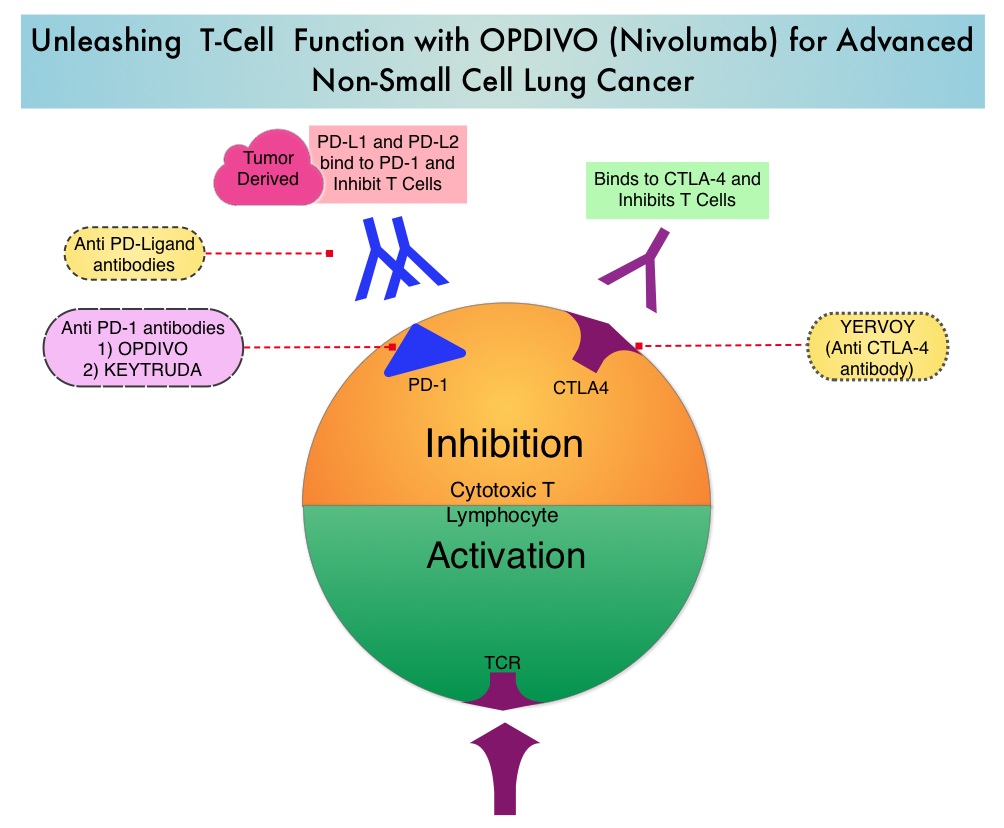
Immune checkpoints are cell surface inhibitory proteins/receptors that are expressed on activated T cells. They harness the immune system and prevent uncontrolled immune reactions. Under normal circumstances, Immune checkpoints or gate keepers inhibit intense immune responses by switching off the T cells of the immune system. With the recognition of Immune checkpoint proteins and their role in suppressing antitumor immunity, antibodies have been developed that target the membrane bound inhibitory Immune checkpoint proteins/receptors such as CTLA-4 (Cytotoxic T-Lymphocyte Antigen 4, also known as CD152), PD-1(Programmed cell Death 1), etc. By doing so, T cells are unleashed, resulting in T cell proliferation, activation and a therapeutic response.Unleashing-T Cell-Function-with-OPDIVO-for-Advanced-Non-Small-Cell-Lung-Cancer

OPDIVO® (Nivolumab) is a fully human, immunoglobulin G4 monoclonal antibody that binds to the PD-1 receptor and blocks its interaction with PD-L1 and PD-L2, thereby undoing PD-1 pathway-mediated inhibition of the immune response and unleashing the T cells. OPDIVO® significantly prolonged Overall Survival (OS) and had a favorable toxicity profile, when compared to TAXOTERE® (Docetaxel), in two open-label, randomized, phase III trials, among patients with advanced squamous (CheckMate 017) or non-squamous (CheckMate 057) NSCLC, who had disease progression during or after platinum-based chemotherapy. This data led to the approval of OPDIVO® in previously treated advanced NSCLC.

There is however limited data on long term efficacy and safety for immune checkpoint inhibitors in patients with NSCLC when compared with chemotherapy. The authors in this publication reported updated efficacy and safety data for OPDIVO® in patients with advanced NSCLC from the CheckMate 017 and CheckMate 057 trials, with a minimum follow up of 2 years in all patients. Patients in both trials had stage IIIB or IV disease and had disease progression during or after platinum-based chemotherapy. In CheckMate 017 study, 272 patients with metastatic squamous NSCLC were randomized to receive OPDIVO® (Nivolumab) 3 mg/kg IV every 2 weeks (N=135) or TAXOTERE® (Docetaxel) 75 mg/m2 IV every 3 weeks (N=137). In CheckMate 057 trial, 582 patients were randomized to receive OPDIVO® 3 mg/kg IV every 2 weeks (N=292) or TAXOTERE® 75 mg/m2 IV every 3 weeks (N=290). Treatment was continued until disease progression or unacceptable toxicity. The minimum follow up for survival was 24.2 months.

The Overall Survival rate at 2 years with OPDIVO® versus TAXOTERE® were 23% vs 8% among squamous NSCLC patients and 29% vs 16% among non-squamous NSCLC patients. The relative reductions in the risk of death with OPDIVO® versus TAXOTERE® was similar to what was reported in the primary analyses. As was reported at the time of the primary analysis of these two trials, the superiority of OPDIVO® over TAXOTERE® was independent of Programmed cell Death Ligand 1 (PD-L1) expression in squamous NSCLC, whereas among patients with non-squamous histology, the benefit with OPDIVO® was greater in those with higher levels of PD-L1 expression, but patients with PD-L1 expression of less than 1% benefited as well.

In the pooled analysis of both trials, the median OS was 11.1 months with OPDIVO® versus 8.1 months with TAXOTERE® (HR=0.72). The relative reduction in the risk of death with OPDIVO® was 28%. Higher PD-L1 expression levels was associated with greater OS benefit with OPDIVO® (HR=0.42 in patients with 50% or more PD-L1 expression). Survival benefit however was still observed in patients with 1% or less PD-L1 expression (HR=0.78). Durable responses were noted with OPDIVO® among 37% of the confirmed responders with squamous NSCLC and 34% of confirmed responders with non-squamous NSCLC. These patients had ongoing responses after a minimum follow up of 2 years whereas no patient in the TAXOTERE® treated group had an ongoing response. The rates of treatment-related adverse events of any grade were lower with OPDIVO® compared to TAXOTERE® (any grade: 68% vs 88%; grade 3-4: 10% vs 55%, respectively).

The authors concluded that OPDIVO® provides long term clinical benefit and has a favorable tolerability profile compared with TAXOTERE®, in previously treated patients with advanced NSCLC. Nivolumab Versus Docetaxel in Previously Treated Patients With Advanced Non–Small-Cell Lung Cancer: Two-Year Outcomes From Two Randomized, Open-Label, Phase III Trials (CheckMate 017 and CheckMate 057). Horn L, Spigel DR, Vokes EE, et al. J Clin Oncol 2017;35:3924-3933

Germline Testing with NGS Multigene Cancer Panels Recommended for All Young Patients with Colorectal Cancer

SUMMARY: ColoRectal Cancer (CRC) is the third most common cancer diagnosed in both men and women in the United States. The American Cancer Society estimates that approximately 140,250 new cases of CRC will be diagnosed in the United States in 2018 and about 50,630 patients are expected to die of the disease. The lifetime risk of developing CRC is about 1 in 21 (4.7%).

Even though the incidence of Colorectal cancer (CRC) in the United States has been rapidly declining overall, primarily driven by screening, the incidence however has been increasing among adults younger than age 50 years. Currently, genetic testing is recommended in patients with polyposis phenotypes, strong family histories, and those whose tumors demonstrate MicroSatellite Instability (MSI) or fail to stain for MisMatch Repair (MMR) gene proteins by immunohistochemistry. Whether patients younger than 50 years of age with CRC, should be tested with a broad multigene panel to detect additional genetic abnormalities, has remained unclear.

The authors conducted this study to ascertain the proportion of young CRC cases associated with genetic predisposition. In this retrospective study, 430 individuals diagnosed with CRC at an age younger than 50 years were evaluated by the clinical genetics service at a single tertiary care cancer center from 1998 through 2015. Data collection included patient histories, tumor phenotypes, and results of germline DNA sequencing. If information was uninformative, germline DNA samples were resequenced using a research-based Next-Generation Sequencing multigene panel. The primary outcome was identification of a pathogenic germline mutation associated with cancer predisposition.

The researchers noted that 26% of young CRC cases had a first-degree relative with CRC and 10% had tumors with histologic evidence for MisMatch Repair (MMR) deficiency and 6% had polyposis phenotype. Germline mutations associated with a hereditary cancer syndrome were identified in 79 patients (18%) following clinically driven germline sequencing, and Next-Generation Sequence analysis using a multigene panel detected actionable germline variants in 6 patients (5%) with uninformative clinical evaluations. Lynch syndrome was identified in 56 patients (14%) of the entire cohort, Familial Adenomatous Polyposis in 2%, MUTYH in 2%, and SMAD4 was mutated in 2 patients, BRCA1 in one, TP53 in one, and CHEK2 in one. Only 51% of the patients with germline mutations associated with a hereditary cancer syndrome gave a family history of CRC diagnosis in a first-degree relative.

The authors concluded that 20% of patients diagnosed with CRC at age younger than 50 years carry a germline mutation associated with cancer, and half of these patients do not have clinical histories typically associated with the identified syndrome. Germline testing with Next-Generation Sequencing multigene cancer panels should therefore be considered for all young patients with CRC. Germline Genetic Features of Young Individuals with Colorectal Cancer. Stoffel EM, Koeppe E, Everett J, et al. Gastroenterology 2017 Nov 13; [e-pub]. (http://dx.doi.org/10.1053/j.gastro.2017.11.004)

Circulating Tumor Cells Predict Late Recurrence in Early Stage HR-Positive Breast Cancer

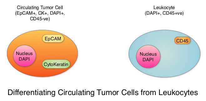
SUMMARY: Breast cancer is the most common cancer among women in the US and about 1 in 8 women (12%) will develop invasive breast cancer during their lifetime. Approximately 266,120 new cases of invasive breast cancer will be diagnosed in 2018 and about 40,920 women will die of the disease. Circulating Tumor Cells (CTCs) are epithelial cells that are shed into the circulation from a primary or metastatic tumor. After being shed, CTCs can remain in the circulation or undergo apoptosis. Evaluation of CTCs during the course of disease has prognostic value. Because of the very low concentrations of CTCs (1 CTC in the background of millions of normal hematopoietic cells) in the peripheral blood, different technologies have been developed that will allow enrichment and detection of these CTCs. One such technology is the CELLSEARCH® system which is the first FDA-approved test for CTC assessment, in the peripheral blood of patients with breast cancer. This automated system is able to enrich the peripheral blood sample with CTCs and the cells then are fluorescently stained for CytoKeratins (CK8,18 and 19), Common Leukocyte Antigen (CD45) and a nuclear dye (DAPI). CTCs are identified when they are CK positive, CD45 negative and DAPI positive. In essence, CTC assessment is a real time, peripheral blood evaluation (“Liquid Biopsy”) in breast cancer patients.Differentiating-Circulating-Tumor-Cells-from-Leukocytes

In women with Hormone Receptor (HR)-positive breast cancer, late recurrences occurring 5 or more years after diagnosis, account for approximately 50% of recurrences. In a meta-analysis from the EBCTCG (Early Breast Cancer Trialists’ Collaborative Group) group, the 10-year risk of recurrence after 5 years of endocrine therapy was found to be 5% in patients with node negative breast cancer, 10% in patients with 1-3 positive lymph nodes, and 22% in patients with 4-9 positive lymph nodes. Late recurrence in patients with HR-positive breast cancer has therefore become a major concern, as this risk for recurrence and death from breast cancer can be ongoing for at least 20 years after the original diagnosis..

The blood and tumor samples of patients are valuable for research and allow the exploration of possible tumor and host-related factors contributing to recurrence. The Coalition of Cancer Cooperative Groups, a non-profit member organization of the National Cancer Institute-sponsored Cooperative Groups, in 2013, created the North American Breast Cancer Groups (NABCG) Biospecimen Bank for Determinants of Late Relapse in Operable Breast Cancer. The purpose for the collection and analysis of samples is, to improve our understanding of breast cancer, and identify which patients are most likely to benefit from specific therapies. The NABCG Biospecimen Bank remains a unique resource of archived primary and metastatic tumor tissue, blood, and DNA, collected from more than 15,000 women, who participated in two large cooperative group cancer treatment trials, TAILORx and E5103.

The authors in this analysis included 546 patients with HER-2-negative, stage II to stage III breast cancer from the E5103 clinical trial, with no clinical evidence of recurrence between 4.5 to 7.5 years following their initial diagnosis. ECOG-ACRIN clinical trial E5103 is a randomized phase III trial, which evaluated the addition of Bevacizumab (AVASTIN®), a Vascular Endothelial Growth Factor (VEGF) inhibitor, to adjuvant chemotherapy following surgery, in patients with lymph node-positive or high-risk, lymph node-negative breast cancer. All patients had undergone surgery and adjuvant chemotherapy as well as endocrine therapy for at least 5 years, if hormone receptor positive. The CTCs from the blood samples of these patients were measured using the CELLSEARCH® CTC assay. None of the patients had clinical evidence of recurrence at the time of this analysis. More than half of the patients (56%) were 50 years or older, 59% of the patients had a tumor 2 cm or larger, 73% had positive lymph nodes and 65% had Hormone Receptor (HR) positive disease. This Primary endpoint of this study was time to recurrence and the primary focus in this analysis was on the group with Hormone Receptor positive disease.

After a median follow up of 1.6 years, 4.8% of patients had a positive CTC assay result overall (1 or more cells/7.5 mL in the blood). Among those with Hormone Receptor positive breast cancer, 5.1% had a positive CTC assay result whereas positive CTC assay result was noted in 4.1% of patients with hormone receptor negative disease. Of the 353 patients with HR-positive disease in this analysis, 4% had a recurrence whereas only 0.5% with HR-negative disease had a local recurrence. The Hazard Ratio for patients with CTC negative results compared with CTC positive results was 21.7 (P<0.001). A positive CTC assay result was associated with a nearly 21.7 fold increased risk of breast cancer recurrence in patients with HR-positive disease. A positive CTC assay was not associated with recurrence in the HR-negative group. The Positive Predictive Value of a positive CTC assay for recurrence at 2 years, in patients with HR-positive disease was 35%, and the Negative Predictive Value for patients in this cohort was 98%.

It was concluded that detection of positive Circulating Tumor Cells in the blood, 5 years after breast cancer diagnosis, was associated with an increased risk for late recurrence, in women with HR-positive, HER2-negative breast cancer. Sparano JA, O’Neill A, Alpaugh K, et al. Circulating tumor cells and late recurrence of breast cancer. Presented at: 2017 San Antonio Breast Cancer Symposium; December 5-9, 2017; San Antonio, TX. Abstract GS6-03.

Monoclonal Gammopathy of Undetermined Significance Associated with Shorter Survival

SUMMARY: Multiple Myeloma (MM) is a clonal disorder of plasma cells in the bone marrow. It evolves from a precursor stage called Monoclonal Gammopathy of Undetermined Significance (MGUS) to MM. Smoldering Multiple Myeloma (SMM) is an intermediate stage in this process of disease evolution. MGUS is defined as the presence of a serum monoclonal protein (M protein) at a concentration of 3 g/dl or less, urine with none or only modest amounts of monoclonal light chains, absence of CRAB features that are related to the M protein (Hypercalcemia, Renal insufficiency, Anemia, and Bone lesions) and 10% or fewer monoclonal plasma cells in the bone marrow. MGUS occurs in approximately 3% of individuals 50 years of age or older and in 5.3% of those 70 years of age or older.

MGUS is classified into two major subtypes, IgM and non-IgM. This is because the clonal cell that is involved and the nature of progression, differ between these two types. The origin of IgM MGUS is typically from a CD20 positive Lymphoplasmacytic cell that has not undergone Switch Recombination (Class-Switch Recombination or CSR, is a biological mechanism that changes a B cell production of immunoglobulin from one type to another, such as from the isotype IgM to the isotype IgG.), and this disease type is associated with a risk of progression to Lymphoma or Waldenström’s macroglobulinemia. In contrast, non-IgM MGUS typically arises from mature plasma cells that have undergone Switch Recombination and is associated with a risk of progression to Multiple Myeloma. Both disease types can progress to AL Amyloidosis.

There is limited information regarding malignant transformation of MGUS to Multiple Myeloma as well as Overall Survival, after diagnosis is made. Further, the risk of progression to Multiple Myeloma among the two major biologic subtypes of MGUS, IgM and non-IgM, is different, and data regarding the prognosis and risk stratification associated with these entities have not been well characterized. The authors in this publication, which is a longer follow up of previously reported findings, studied 1384 patients with MGUS, who were identified in a well-defined geographic area, from 1960-1994. The median follow up was 34.1 years. The Primary end point was progression to Multiple Myeloma or another Plasma-cell or Lymphoid disorder.

During 14,130 person-years of follow up, the rate of progression of MGUS to Multiple Myeloma was 6.5 times as high as the rate in the control population. The risk of progression to Multiple Myeloma was 10% at 10 years, 18% at 20 years, 28% at 30 years, 36% at 35 years, and 36% at 40 years. The risk of progression varied depending on the biologic subtypes of MGUS and other adverse risk factors. Among patients with IgM MGUS, the presence of two adverse risk factors – an abnormal serum free light chain ratio (ratio of kappa to lambda free light chains) and a high serum monoclonal protein level (1.5 g/dl or greater), was associated with a risk of progression at 20 years of 55%, as compared with 41% among patients who had one adverse risk factor and 19% among patients who had neither risk factor. Among patients with non-IgM MGUS, the risk of progression to Multiple Myeloma at 20 years was 30% among those who had the two risk factors, 20% among those who had one risk factor, and 7% among those who had neither risk factor. More importantly, patients with MGUS had shorter survival compared to age and sex matched control population (median 8.1 versus 12.4 years, P<0.001).

It was concluded that there were significant differences noted in the risk of progression between patients with IgM MGUS and those with non-IgM MGUS and Overall Survival was shorter among patients with MGUS than was expected in a matched control population. Long-Term Follow-up of Monoclonal Gammopathy of Undetermined Significance. Kyle RA, Larson DR, Therneau TM, et al. N Engl J Med 2018; 378:241-249